. 2007 Sep;37(3):273-90.
doi: 10.1165/rcmb.2005-0460OC. Epub 2007 Apr 26.

Central role of Muc5ac expression in mucous metaplasia and its regulation by conserved 5' elements

Affiliations

Central role of Muc5ac expression in mucous metaplasia and its regulation by conserved 5' elements

Hays W J Young et al. Am J Respir Cell Mol Biol. 2007 Sep.

Abstract

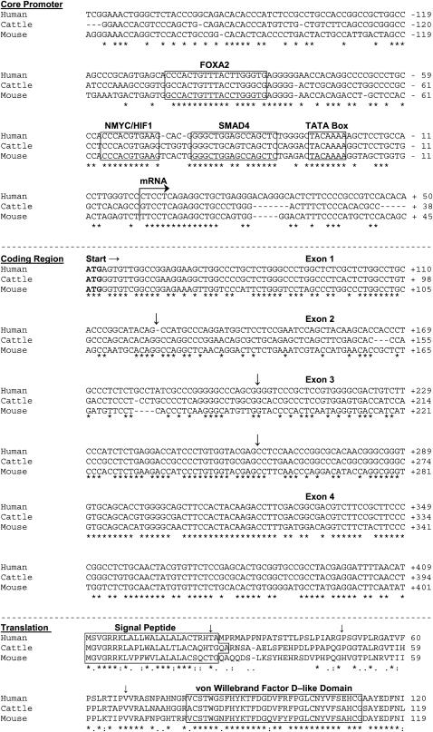
Mucus hypersecretion contributes to morbidity and mortality in many obstructive lung diseases. Gel-forming mucins are the chief glycoprotein components of airway mucus, and elevated expression of these during mucous metaplasia precedes the hypersecretory phenotype. Five orthologous genes (MUC2, MUC5AC, MUC5B, MUC6, and MUC19) encode the mammalian gel-forming mucin family, and several have been implicated in asthma, cystic fibrosis, and chronic obstructive pulmonary disease pathologies. However, in the absence of a comprehensive analysis, their relative contributions remain unclear. Here, we assess the expression of the entire gel-forming mucin gene family in allergic mouse airways and show that Muc5ac is the predominant gel-forming mucin induced. We previously showed that the induction of mucous metaplasia in ovalbumin-sensitized and -challenged mouse lungs occurs within bronchial Clara cells. The temporal induction and localization of Muc5ac transcripts correlate with the induced expression and localization of mucin glycoproteins in bronchial airways. To better understand the tight regulation of Muc5ac expression, we analyzed all available 5'-flanking sequences of mammalian MUC5AC orthologs and identified evolutionarily conserved regions within domains proximal to the mRNA coding region. Analysis of luciferase reporter gene activity in a mouse transformed Clara cell line demonstrates that this region possesses strong promoter activity and harbors multiple conserved transcription factor-binding motifs. In particular, SMAD4 and HIF-1alpha bind to the promoter, and mutation of their recognition motifs abolishes promoter function. In conclusion, Muc5ac expression is the central event in antigen-induced mucous metaplasia, and phylogenetically conserved 5' noncoding domains control its regulation.


Figures

<b>Figure 1.</b>
Figure 1.
A single antigen challenge causes goblet cell metaplasia and alters mucin gene expression in sensitized mice. (A) The airway epithelium of saline challenged mice contains few or no PAFS-positive mucin granules. (B) Three days after antigen exposure, the airways demonstrate an increase in PAFS-positive mucin granules (red). (C) RT-PCR analysis of gel-forming mucin genes over a 1-wk time course of goblet cell metaplasia after a single antigen challenge demonstrates that Muc5ac, Muc5b, and scant Muc19 are detectable at baseline and after antigen challenge while no Muc2 or Muc6 are detectable at any time points. Magnification bar = 50 μm for A and B. Positive controls in C are PCR of reverse-transcribed total RNA extracted from colon (Muc2, 1,046 bp), stomach (Muc5ac, 414 bp and Muc6, 791 bp), tongue (Muc5b, 512 bp), and salivary gland (Muc19, 451 bp). Data are single representatives of samples from 5–12 mice per group.
<b>Figure 2.</b>
Figure 2.
Muc5ac is selectively induced in the lungs of antigen-challenged mice. (A) Quantitative RT-PCR of total lung RNA demonstrates predominant Muc5b mRNA at baseline (open bars). Muc5ac mRNA is present at ∼ 30-fold lower levels than Muc5b while Muc2 and Muc19 mRNAs are present at ∼ 300 and > 700-fold lower levels than Muc5b, respectively. Three days after antigen challenge (filled bars), Muc5ac is the only gel-forming mucin whose mRNA levels increase significantly. (B) The magnitude of Muc5ac induction, compared with its expression level in saline-challenged mice (S), is significant 72 h after exposure, but not at earlier time points. Muc5ac (upright triangles) induction is also of significantly greater magnitude than any changes seen in Muc2 (squares), Muc5b (inverted triangles), or Muc19 (diamonds) mRNA levels at any time points studied here. Data are presented as means ± SE of triplicate samples from 5–12 mice per group. *Significant difference from other mucin mRNAs within a given treatment group/time point. #Significant difference between an individual mucin and its baseline expression level. Note Log2 scale for y axis in A.
<b>Figure 3.</b>
Figure 3.
Coordinate expression of Muc5ac mRNA and PAFS-positive mucin staining within the bronchial epithelium of antigen-challenged mice three days after single antigen challenge. In situ hybridization was used to detect Muc5ac mRNA in lung sections from antigen-challenged (A–D) and saline-challenged (G and H) mice. The antisense Muc5ac riboprobe hybridizes to the epithelium in the bronchial (Br) airways (red in A and C), but not in the bronchiolar (br) airways (A and D). PAFS staining of a consecutive airway section shows mucin (red in E) in the same cells of the bronchial airways that show Muc5ac mRNA positivity in A and C. No PAFS-positive mucin staining is present within epithelium of the bronchiolar airways (F) that also showed no Muc5ac mRNA (D). Lack of signal detection after incubation with sense riboprobe in the opposite consecutive antigen-challenged lung section as that shown in E and F (B) or incubation with antisense riboprobe in lung sections of saline challenged animals demonstrates specificity of the findings in A, C, and D. Magnification bar = 100 μm. Blue in A–D, G, and H is Hoechst 33258. Green in E and F is intercalated acriflavine staining of nuclear and cytoplasmic nucleic acids.
<b>Figure 4.</b>
Figure 4.
Alignment of 5′ noncoding, coding, and translated sequence fragments of mammalian MUC5AC orthologs. (Top section) The ∼ 200-bp segment of the 5′ flanking region proximal to the conserved transcriptional initiation site (arrow) shows 55% cross-species identity and conserved consensus sequences for TFs (boxed segments). (Middle section) The ∼ 360 bp four exon amino terminal coding cDNA segments show 65% cross-species identity. (Bottom section) Translation of the four-exon segment above shows a conserved signal peptide and a highly conserved von Willebrand Factor D–like Domain that is a characteristic of the amino termini of all gel-forming mucins. Inverted arrows depict exon junctions.
<b>Figure 5.</b>
Figure 5.
Evolutionary conservation of the 5′ flanking region of mouse and human MUC5AC orthologs. (Top) Evolutionarily conserved regions (ECRs) within the MUC2–MUC5AC intergene sequences of mice (black) and humans (red) were identified using BLASTz alignment to show the presence of discrete blocks of ECRs. ECRs are most frequent in the proximal promoter-enhancer regions (∼ 7 kb in humans and ∼ 5 kb in mice). (Bottom) rVISTA analysis shows conservation of TF consensus sequences in mouse (black) and human (red) proximal promoters. Numbers below axes depict distance (bp) from transcriptional start site.
<b>Figure 6.</b>
Figure 6.
Mouse transformed Clara cells express markers of mucous metaplasia. (A) Cultures of mtCC1–2s were serum starved for 6 h and then incubated with IL-13 (100 ng/ml; black bars) or EGF (25 ng/ml; gray bars) for 24 h. Quantitative RT-PCR was then used to assess Muc2, Muc5ac, Muc5b, and Muc19 mucin mRNAs. CCSP and Clca3 were also assessed as independent markers of baseline and mucous metaplastic gene expression, respectively. Muc5ac, CCSP, and Clca3 (but not Muc2, Muc5b, or Muc19) are significantly increased by IL-13. Only Muc5ac and Clca3 are significantly increased by EGF in mtCC1–2s. Data are means ± SE from three independent experiments conducted in triplicate. (B) PAFS staining of a tissue section from a 20-wk-old CCSP-SV40 T Ag transgenic mouse demonstrates the presence of bronchiolar tumors (white asterisks), some of which contain mucous cells (box in B shown at higher magnification image in C). Arrow in C identifies a region of PAFS-positive papillary tumor growth shown in the inset. Magnification bar = 150 μm (B), 30 μm (C), and 10 μm (inset). Note Log2 scale for y axis in A. *Significant difference between an individual mRNA and its baseline expression level.
<b>Figure 7.</b>
Figure 7.
Muc5ac promoter activity in mouse transformed Clara cells. 1–5 kb Muc5ac promoter-luciferase constructs were transfected into mtCC1–2's (black bars) and 3T3 cells (white bars) and incubated overnight in serum supplemented medium. The 1 and 2 kb domains proximal to the translation start site are strongly activated in mtCC1–2s, but not 3T3 cells. By contrast, inclusion of distal domains (3–5 kb upstream) abolishes luciferase activity. Like the Muc5ac promoter, the 800-bp core promoter of CCSP also directs Clara cell–selective transcriptional activation. Dashed line is luciferase activity in cells transfected with empty pGL3 vector. Data are presented as means ± SE of triplicate samples from five experiments. *Significant difference from 3T3 cells, and #significant difference from 1 kb promoter activity in mtCC1–2s.
<b>Figure 8.</b>
Figure 8.
The third (−2/−3) kb domain of the 5′ flanking region of Muc5ac contains repressor(s) that act redundantly upon Clara cell secretory products. (Top) mtCC1–2s were transfected with constructs containing either CMV (white bar) or CCSP (black bar) promoter driven luciferase and incubated overnight in serum supplemented medium, and these direct high levels of luciferase activity. (Bottom) Chimeric promoter constructs in which the −2/−3 kb repressor domain of the Muc5ac 5′ flanking region is present 5′ to CMV (white bar) or CCSP (black bar) promoter driven luciferase show uninhibited CMV-driven luciferase activity and ablated CCSP promoter-driven luciferase activity. Data are presented as means ± SE of triplicate samples from three experiments. *Significant difference from −2/−3 kb CMV, and nonchimeric CMV and CCSP promoter–driven reporters.
<b>Figure 9.</b>
Figure 9.
Muc5ac promoter activity is controlled by functional evolutionarily conserved cis elements. Site-directed mutagenesis was used to disrupt TF motifs contained within the −1 kb Muc5ac firefly luciferase construct used in Figure 7. Cultures were transfected with 1 μg of DNA and incubated in the presence of 10% fetal bovine serum (FBS) to maximally activate promoter activity overnight. Luciferase and BCA assays were then performed. Data are presented as the percentage of activity of mutated promoters compared with wild type. Mutation of individual Hif1/Nmyc, Smad4, and Lef consensus sites identified in silico significantly impairs Muc5ac promoter function in vitro. Combined mutation of Hif1/Nmyc and Smad4 sites abolishes promoter activity. Dashed line represents baseline wild-type 1-kb Muc5ac promoter activity in the absence of FBS. Values are means ± SE of results from three separate transfection experiments performed in sextuplicate. *Significant difference from wild type.
<b>Figure 10.</b>
Figure 10.
Activation of the Muc5ac promoter is cytokine inducible. Muc5ac promoter–luciferase constructs (1–5 kb) were transfected into mtCC1–2s and incubated overnight in serum-supplemented medium. Cells were serum starved for 6 h, followed by overnight incubation with SFM alone (open bars), 100 ng/ml recombinant mouse IL-13 (solid bars), or 25 ng/ml recombinant mouse EGF (shaded bars). The Muc5ac 5′ flanking region is differentially regulated in mtCC1–2s after IL-13 or EGF stimulation as compared with SFM (open bars). The 800-bp CCSP core promoter is unresponsive to IL-13 and EGF. Dashed line is luciferase activity in cells transfected with empty pGL3 vector. Data are presented as means ± SE in triplicate samples in a representative of six experiments. *Significant difference from medium alone.
<b>Figure 11.</b>
Figure 11.
Cytokine inducible Muc5ac promoter activity is mediated by cis SMAD4 and HIF-1/Nmyc recognition motifs. Cultures of mtCC1–2s were transfected with 1 μg of wild-type and the SMAD4 and HIF-1/Nmyc mutant −1 kb Muc5ac firefly luciferase constructs used in Figure 9 and incubated in the presence of 10% FBS overnight. The following day, cells were serum starved for 6 h and were incubated overnight in SFM alone (open bars), SFM supplemented with 100 ng/ml recombinant mouse IL-13 (solid bars in A), or SFM supplemented with 25 ng/ml recombinant mouse EGF (solid bars in B). Eighteen hours later, luciferase and BCA assays were performed. In cells transfected with the wild-type promoter IL-13 and EGF, both increase Muc5ac promoter activity compared with unstimulated cells. In cells transfected with the −1 kb promoter harboring mutant HIF or SMAD4 motifs, singly and in combination, IL-13 and EGF both fail to induce promoter activity. Data are presented as luciferase activity normalized to total protein within each well. Values are means ± SE of results from three separate transfection experiments performed in sextuplicate. *Significant difference from wild-type promoter-transfected cells cultured in SFM.
<b>Figure 12.</b>
Figure 12.
HIF-1α and SMAD4 bind to the Muc5ac promoter in mouse transformed Clara cells. Cells were cultured in the presence of 10% FBS until 75–90% confluent. Cells were then serum-starved for 6 h and were incubated overnight in SFM alone, SFM supplemented with 100 ng/ml recombinant mouse IL-13, or SFM supplemented with 25 ng/ml recombinant mouse EGF. Eighteen hours later, cells were fixed and lysed for chromatin immunoprecipitation (ChIP) using HIF-1α and SMAD4 Abs. (A) Nonquantitative PCR analysis of precipitated and input DNA using Muc5ac promoter–specific primers that flank the SMAD4 and HIF-1α TF motifs (sense primer, −144 to −125 bp ; antisense primer +20 to +1; see Figure 4) demonstrates binding of HIF-1α and SMAD4 to the Muc5ac promoter at baseline and after cytokine stimulation. (B) Quantitative PCR analysis of precipitated DNA demonstrates that HIF-1α and SMAD4 both bind to the Muc5ac promoter at baseline, and SMAD4 binds ∼ 3 times more often. HIF-1α binding to the Muc5ac promoter increases in cytokine stimulated cells, but SMAD4 binding decreases. Assessment of DNA precipitated with control IgG demonstrated ∼ 10,000 to > 100,000-fold lower values than any of the values generated from PCR of DNA precipitated by SMAD4 and HIF-1α Abs. Template DNA was diluted 10-fold for PCR analysis of input in A. Values in B are means ± SE of results normalized to input from three separate ChIP experiments analyzed in triplicate. *Significant difference in HIF-1α and SMAD4 association with the Muc5ac promoter between cytokine stimulated cells and cells cultured in SFM. #Significant difference between SMAD4 and HIF-1α results in cells cultured under identical conditions.

References

    1. Wanner A, Salathe M, O'Riordan TG. Mucociliary clearance in the airways. Am J Respir Crit Care Med 1996;154:1868–1902. - PubMed
    1. Evans CM, Agrawal A, Dickey BF. Strategies to reduce excessive mucus secretion in airway inflammation. Eissa T, Huston D, editors. Therapeutic targets of airway inflammation. New York: Marcel Dekker; 2002.
    1. Williams OW, Sharafkhaneh A, Kim V, Dickey BF, Evans CM. Airway mucus: from production to secretion. Am J Respir Cell Mol Biol 2006;34:527–536. - PMC - PubMed
    1. Boers JE, Ambergen AW, Thunnissen FB. Number and proliferation of clara cells in normal human airway epithelium. Am J Respir Crit Care Med 1999;159:1585–1591. - PubMed
    1. Evans CM, Williams OW, Tuvim MJ, Nigam R, Mixides GP, Blackburn MR, DeMayo FJ, Burns AR, Smith C, Reynolds SD, et al. Mucin is produced by clara cells in the proximal airways of antigen-challenged mice. Am J Respir Cell Mol Biol 2004;31:382–394. - PMC - PubMed

Publication types

MeSH terms