. 2007 May;153(Pt 5):1593-1603.
doi: 10.1099/mic.0.2006/004200-0.

The outer membrane secretin PilQ from Neisseria meningitidis binds DNA

Affiliations

The outer membrane secretin PilQ from Neisseria meningitidis binds DNA

Reza Assalkhou et al. Microbiology (Reading). 2007 May.

Abstract

Neisseria meningitidis is naturally competent for transformation throughout its growth cycle. Transformation in neisserial species is coupled to the expression of type IV pili, which are present on the cell surface as bundled filamentous appendages, and are assembled, extruded and retracted by the pilus biogenesis components. During the initial phase of the transformation process, binding and uptake of DNA takes place with entry through a presumed outer-membrane channel into the periplasm. This study showed that DNA associates only weakly with purified pili, but binds significantly to the PilQ complex isolated directly from meningococcal membranes. By assessing the DNA-binding activity of the native complex PilQ, as well as recombinant truncated PilQ monomers, it was shown that the N-terminal region of PilQ is involved in the interaction with DNA. It was evident that the binding of ssDNA to PilQ had a higher affinity than the binding of dsDNA. The binding of DNA to PilQ did not, however, depend on the presence of the neisserial DNA-uptake sequence. It is suggested that transforming DNA is introduced into the cell through the outer-membrane channel formed by the PilQ complex, and that DNA uptake occurs by non-specific introduction of DNA coupled to pilus retraction, followed by presentation to DNA-binding component(s), including PilQ.


Figures

Fig. 1.
Fig. 1.
Detection of pilus–DNA interaction in solution. DNA was immobilized in the well and overlaid with protein in solution. (a) Binding of DNA glycosylase MutY to DNA (positive control protein). Saturation was reached at 1 μg MutY. One representative experiment out of four is shown. (b) Binding of N. meningitidis M1080 and P. aeruginosa PAK pili to DNA. The mean of four independent measurements using each purified pilus preparation is shown. Error bars indicate sd. (c) Competition of pili binding to immobilized DNA by preincubation with DNA in solution. The average of two independent measurements using pilus protein is shown, as above. Error bars indicate minimum and maximum values. (d) Competition of pili binding to immobilized DNA by preincubation with pilus-specific antibodies. The average of two independent measurements using 0.125 μl pilus proteins is shown, as above. Error bars indicate minimum and maximum values.
Fig. 2.
Fig. 2.
Gene organization and pilQ constructs. (a) Schematic representation of the N. meningitidis strain M1080 pilQ gene and the predicted gene product. The positions of the N-terminal SBRs and the PstI restriction site are indicated. The locations of positively charged areas within the predicted sequence are marked in black. (b) Schematic representation of the PilQ truncated proteins used in this study, depicted relative to Fig. 2(a). The position of the hexa-histidine tag is indicated with an ‘H’ and the sizes of the recombinant proteins are given on the right.
Fig. 3.
Fig. 3.
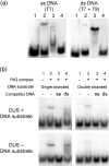
DNA-binding characteristics of the native PilQ complex measured by band-shift analysis. The interaction of native PilQ complex with ssDNA and dsDNA substrates was studied. (a) Band-shift analysis of PilQ complex binding to ssDNA (T7) or dsDNA (T7+T8 hybrid) substrates. Lane 1, free labelled DNA substrates (no PilQ); lane 2, DNA and PilQ complex; lane 3, DNA and Taq polymerase (positive control); lane 4, DNA and BSA (negative control). (b) Competition band-shift experiments, showing the ability of unlabelled ss- or dsDNA ligands to compete with the binding of labelled ligands. Results are compared for oligonucleotides containing (T1/T2) or not containing (T3/T4) DUSs. Lane 1, free labelled DNA substrates; lanes 2–4, PilQ complex.
Fig. 4.
Fig. 4.
Binding of the PilQ complex to DNA assessed by a solid-phase overlay assay in the form of Southwestern analysis. Southwestern analysis of the proteins reacted with ssDNA substrate (T1) (a) and immunoblotting with PilQ-specific antibody (b) is shown. Lanes 1, M1080 whole-cell lysate; lanes 2, M1080-ΔPilQ whole-cell lysate; lanes 3, MutY (positive control); lanes 4, BSA (negative control). Markers on the left give the molecular mass in kDa; arrows on the right indicate the migration positions of the PilQ complex, PilQ monomer, PilQ degradation product typical for secretins, and DNA glycosylase MutY (positive control protein for DNA binding).
Fig. 5.
Fig. 5.
Interaction of recombinant PilQ with DNA substrates in band-shift analysis: dependence on protein concentration and DNA substrate. Free labelled (2000 c.p.m.) ssDNA T7 and dsDNA (T7+T8 hybrid) ligands were incubated with the indicated amount of recombinant N-terminal PilQ protein PilQ25–354, and run under standard conditions. The migration of the shifted DNA substrates appeared to be affected by the PilQ protein concentration and/or charge. Filled arrow, free oligonucleotide; open arrow, partial shift, typical of PilQ25–354; filled arrowhead, full band shift. At high protein concentrations, the sticking of oligonucleotides to protein in the well was also observed.
Fig. 6.
Fig. 6.
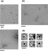
Interaction between DNA and the PilQ complex visualized by transmission electron microscopy. (a) PilQ complex incubated with avidin–nanogold plus biotinylated dsDNA; complexes are indicated with arrows. Bar, 50 nm. (b) PilQ complex incubated with avidin–nanogold plus biotinylated ssDNA; complexes are indicated with arrows. Bar, 50 nm. (c) PilQ complex incubated with avidin–nanogold; nanogold particles alone are indicated by white arrows, and an unliganded PilQ particle is indicated by the black arrow (negative control). Bar, 50 nm. (d) Panel of example particles in each class: left column, PilQ–avidin–nanogold plus biotinylated ssDNA; middle column, unlabelled PilQ; right column, avidin–nanogold label alone. The perimeter of each particle is indicated. Bar, 16.5 nm. For clarity, the contrast in all the figures has been enhanced.

References

    1. Aas, F. E., Wolfgang, M., Frye, S., Dunham, S., Lovold, C. & Koomey, M. (2002). Competence for natural transformation in Neisseria gonorrhoeae: components of DNA binding and uptake linked to type IV pilus expression. Mol Microbiol 46, 749–760. - PubMed
    1. Bille, E., Zahar, J. R., Perrin, A., Morelle, S., Kriz, P., Jolley, K. A., Maiden, M. C., Dervin, C., Nassif, X. & Tinsley, C. R. (2005). A chromosomally integrated bacteriophage in invasive meningococci. J Exp Med 201, 1905–1913. - PMC - PubMed
    1. Bitter, W., Koster, M., Latijnhouwers, M., de Cock, H. & Tommassen, J. (1998). Formation of oligomeric rings by XcpQ and PilQ, which are involved in protein transport across the outer membrane of Pseudomonas aeruginosa. Mol Microbiol 27, 209–219. - PubMed
    1. Bøvre, K., Bergan, T. & Froholm, L. O. (1970). Electron microscopical and serological characteristics associated with colony type in Moraxella nonliquefaciens. Acta Pathol Microbiol Scand [B] Microbiol Immunol 78, 765–779. - PubMed
    1. Bradford, M. M. (1976). A rapid and sensitive method for the quantitation of microgram quantities of protein utilizing the principle of protein-dye binding. Anal Biochem 72, 248–254. - PubMed

Publication types

MeSH terms

LinkOut - more resources