. 2007 May 16;26(10):2552-61.
doi: 10.1038/sj.emboj.7601700. Epub 2007 Apr 26.

Autoprocessing of the Vibrio cholerae RTX toxin by the cysteine protease domain

Affiliations

Autoprocessing of the Vibrio cholerae RTX toxin by the cysteine protease domain

Kerri-Lynn Sheahan et al. EMBO J. .

Abstract

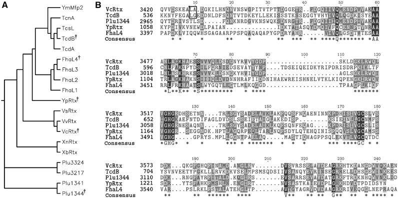
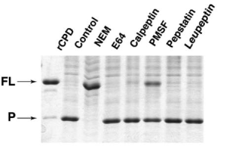
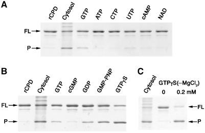
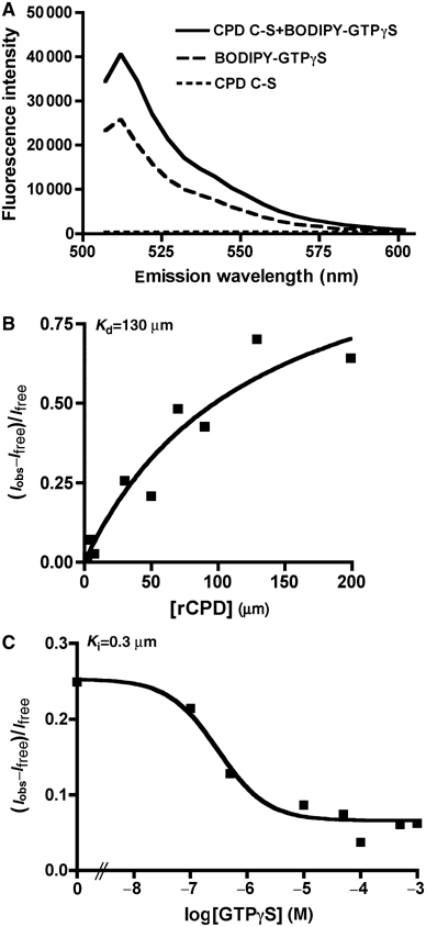
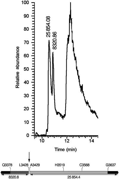
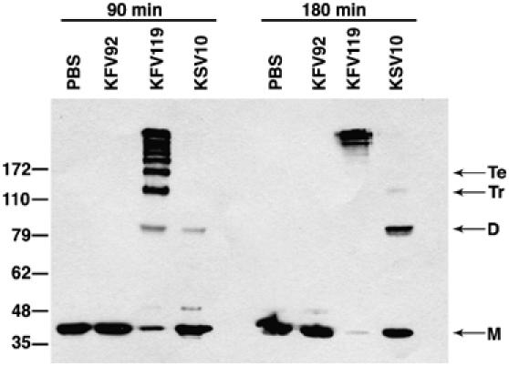
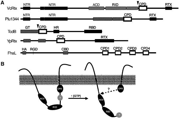
Vibrio cholerae RTX is a large multifunctional bacterial toxin that causes actin crosslinking. Due to its size, it was predicted to undergo proteolytic cleavage during translocation into host cells to deliver activity domains to the cytosol. In this study, we identified a domain within the RTX toxin that is conserved in large clostridial glucosylating toxins TcdB, TcdA, TcnA, and TcsL; putative toxins from V. vulnificus, Yersinia sp., Photorhabdus sp., and Xenorhabdus sp.; and a filamentous/hemagglutinin-like protein FhaL from Bordetella sp. In vivo transfection studies and in vitro characterization of purified recombinant protein revealed that this domain from the V. cholerae RTX toxin is an autoprocessing cysteine protease whose activity is stimulated by the intracellular environment. A cysteine point mutation within the RTX holotoxin attenuated actin crosslinking activity suggesting that processing of the toxin is an important step in toxin translocation. Overall, we have uncovered a new mechanism by which large bacterial toxins and proteins deliver catalytic activities to the eukaryotic cell cytosol by autoprocessing after translocation.


Figures

Figure 1
Figure 1
Alignment of putative cysteine protease domains. (A) Phylogenetic tree of 19 putative CPDs and (B) CLUSTALW alignment of five diverse sequences. Symbol in phylogenetic tree indicates diverse sequences selected for alignment in (B). In (B), asterisks in consensus sequence represent conserved residues in 3/5 sequences and capital letters represent 100% identity. Outlined dipeptide sequence indicates cleavage sites determined experimentally. CPDs were identified within nine Vibrio-type RTX toxins from V. cholerae (VcRtx); V. vulnificus (VvRtx), V. splendidus (VsRtx), Xenorhabdus nematophila (XnRtx), X. bovienii (XbRtx), and Photorhabdus luminescens (Plu1344, Plu1341, Plu3217, and Plu3324); four clostridial toxins, specifically C. difficile toxin A (TcdA), toxin B (TcdB), C. sordellii cytotoxin L (TcsL), and C. noveyi alpha toxin (TcnA); two putative Yersinia toxins Y. pseudotuberculosis YPTB3219 (YpRtx) and Y. mollaretti Mfp2 (YmMfp2); and four domains arranged in tandem in B. pertussis putative adhesin FhaL (FhaL1-4).
Figure 2
Figure 2
Expression of RTX amino acids 3376–3637 is cytotoxic to cells and the protein is processed intracellularly. COS-7 cells transiently expressing EGFP (A), CPDc-EGFP (B), CPDc C-S-EGFP (C), and CPDc H-A-EGFP (D) were observed by fluorescence microscopy. Representative images are displayed as an overlay of the fluorescence micrograph obtained at 550–575 nm to detect GFP and 440–470 nm to detect Hoechst staining. (E) At 24 h after transfection, cells were resuspended in SDS buffer, boiled, and subjected to SDS–PAGE and Western blotting with an anti-GFP antibody.
Figure 3
Figure 3
Transiently expressed CPD in cells is autoprocessed within the N-terminus. (A) Schematic representation of fusion proteins with the predicted mw of each full-length (FL) fusion protein indicated at right. Numbers along the bottom correspond to the amino-acid sequence of translated products according to the RTX toxin annotation of Lin et al (1999) (GenBank accession no. gi ∣4455065). (B) Expression of the CPD and CPD C-S fusion proteins was detected in transiently transfected COS-7 cells by Western blotting for GFP. Arrows mark full-length (FL) and the processed (P) forms of CPD.
Figure 4
Figure 4
rCPD undergoes in vitro processing after addition of host cell lysate. (A) Schematic representation of rCPD (gray shading) with N- and C-terminal 6 × His tag fusions (black shading). (B–E) In vitro processing reactions were performed with 2 μg rCPD incubated in the presence of lysate and samples were separated by SDS–PAGE and stained with Coomassie R250. (B) rCPD was incubated with 5 μg of total cell protein and the reaction was stopped at the indicated time points. (C) Overnight incubation of rCPD with increasing amounts of lysate as indicated. (D) rCPD was incubated with equivalent volumes of lysate, boiled lysate, or lysate pretreated for 15 min with 200 μg/ml of Proteinase K, DNase, or RNase. rCPD was also incubated with membrane and cytosolic fractions (5 μg of total cell protein) obtained from subcellular fractionation of the lysate. (E) rCPD and rCPD C-S were incubated for 2 h in the absence (lanes 1 and 3) or presence of 5 μg of total cell protein (lanes 2 and 4). Arrows mark full-length (FL) and the processed (P) forms of rCPD.
Figure 5
Figure 5
Autoprocessing of rCPD is inhibited by NEM. rCPD (2 μg) was preincubated for 30 min with 1 mM NEM, E-64, Calpeptin, or PMSF, 100 μM Pepstatin, or 200 μM Leupeptin. Then, 5 μg of total cell protein was added to stimulate processing followed by overnight incubation. Proteins were separated by SDS–PAGE and stained with Coomassie R250. Arrows mark full-length (FL) and the processed (P) forms of rCPD.
Figure 6
Figure 6
rCPD processing is stimulated by guanine nucleotides. (A, B) rCPD (2 μg) was incubated alone (rCPD), with 5 μg of cytosolic protein preparation (Cytosol), or with 5 mM MgCl2 plus 5 mM of the indicated compound. (C) Incubation of 2 μg rCPD with the indicated concentration of GTPγS in the absence of MgCl2. All reactions were terminated after 2 h at 37°C. Proteins were separated by SDS–PAGE and stained with Coomassie R250. Arrows mark full-length (FL) and the processed (P) forms of rCPD.
Figure 7
Figure 7
rCPD C-S binds GTPγS. (A) Representative emission scan after excitation at 488 nm of 200 μM rCPD C-S alone, 0.25 μM BODIPY FL- GTPγS alone, or rCPD mixed with BODIPY FL- GTPγS showing maximum fluorescence intensity at 512 nm and the absence of background fluorescence from the protein. (B) Binding curve of varying concentration of rCPD incubated with 0.25 μM BODIPY FL- GTPγS. (C) Competitive inhibition curve of 50 μM CPD incubated with 0.25 μM BODIPY FL- GTPγS with varying concentrations of unlabeled GTPγS. Kd and Ki were determined as described in Materials and methods.
Figure 8
Figure 8
rCPD is cleaved between L3428 and A3429. Processed rCPD eluted from C8 reverse phase column in two peaks at 10.56 and 10.88 min. The third peak at 12.31 min consists of poorly resolved materials such a polymers, small molecules, and peptides. The mass of the proteins contained within the peptide fragment peaks was determined by LTQ-FT analysis and the results are shown above the peak. Schematic representation of rCPD shows the determined cleavage site as well as the computer-predicted mass of the two fragments.
Figure 9
Figure 9
Cys3568 is important for RTX toxin actin crosslinking activity. COS-7 cells were incubated with PBS or V. cholerae strains with an intact rtxA gene (KFV119), a null mutation in rtxA (KFV92), or an rtxA gene with a C3568S point mutation (KSV10). Cells were harvested after 90 and 180 min of incubation, and actin crosslinking was measured by Western blotting with an anti-actin antibody. Lines at right mark monomer (M), dimer (D), trimer (Tr), and tetramer (Te) forms of actin.
Figure 10
Figure 10
Autoprocessing by CPD is a common mechanism for processing large bacterial toxins. (A) Schematic representation of CPD location within large bacterial proteins. CPD is marked as an outlined open box. Black boxes represent regions predicted to be critical for toxin entry (NTR, N-terminal repeats; RTX, RTX repeats; HR, hydrophobic region; RBD, receptor binding domain). Grey hatched boxes represent known or predicted functional regions (ACD, actin crosslinking domain; GT, glucosyltransferase; HA, hemagglutinin; CBD, carbohydrate binding domain; RGD, integrin binding; RID, Rho-inactivation domain). Arrowheads mark cleavage sites determined experimentally. (B) Proposed model of RTX toxin processing.

References

    1. Altschul SF, Madden TL, Schaffer AA, Zhang J, Zhang Z, Miller W, Lipman DJ (1997) Gapped BLAST and PSI-BLAST: a new generation of protein database search programs. Nucleic Acids Res 25: 3389–3402 - PMC - PubMed
    1. Anderson BM, Vasini EC (1970) Nonpolar effects in reactions of the sulfhydryl group of papain. Biochemistry 9: 3348–3352 - PubMed
    1. Bailey TL, Elkan C (1994) Fitting a mixture model by expectation maximization to discover motifs in bioploymers. In Proceedings of the Second International Conference on Intelligent Systems for Molecular Biology, pp 28–36. Menlo Park, CA: AAAI Press - PubMed
    1. Bailey TL, Gribskov M (1998) Combining evidence using p-values: application to sequence homology searches. Bioinformatics 14: 48–54 - PubMed
    1. Barroso LA, Moncrief JS, Lyerly DM, Wilkins TD (1994) Mutagenesis of the Clostridium difficile toxin B gene and effect on cytotoxic activity. Microb Pathog 16: 297–303 - PubMed

Publication types

MeSH terms

LinkOut - more resources