

The quantitative measurements of the intervertebral angulation and translation during cervical flexion and extension
- PMID: 17464516
- PMCID: PMC2200749
- DOI: 10.1007/s00586-007-0372-4
The quantitative measurements of the intervertebral angulation and translation during cervical flexion and extension
Abstract
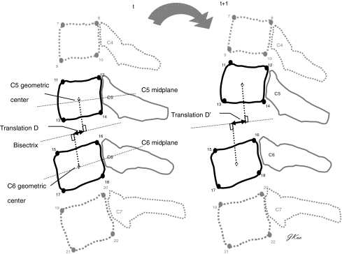
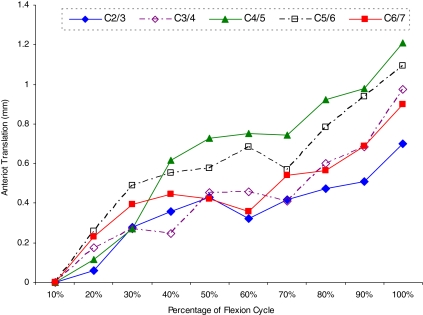
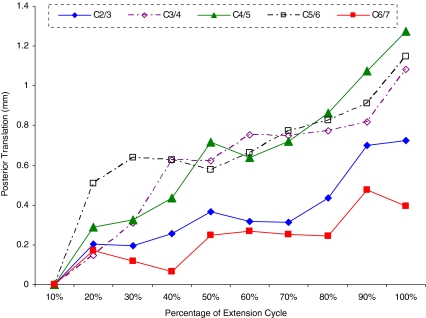
The insufficient exploration of intervertebral translation during flexion and extension prevents the further understanding of the cervical biomechanics and treating the cervical related dysfunction. The objective of this study was to quantitatively measure the continuous intervertebral translation of healthy cervical spine during flexion and extension by videofluoroscopic technique. A total of 1,120 image sequences were analyzed for 56 healthy adult subjects by a precise image protocol during cervical flexion and extension. O: ur results showed there were no statistical angular differences among five spinal levels in either flexion or extension, except for the comparison between C2/3 (13.5 degrees) and C4/5 (22.6 degrees) angles. During cervical flexion, the smallest anterior translations were 0.7 mm at C2/3 level, followed by 0.9 mm at C6/7, 1.0 mm at C3/4, 1.1 mm at C5/6, and the largest 1.2 mm at C4/5 levels. The significantly greater translations were measured in the posterior direction at C3/4 (1.1 mm, P = 0.037), C4/5 (1.3 mm, P = 0.039), and C5/6 (1.2 mm, P = 0.005) levels than in the anterior one. The relatively fluctuant and small average posterior translation fashion at C6/7 level (0.4 mm) possibly originated from the variations in the direction of translation during cervical extension among subjects. Normalization with respect to the widths of individual vertebrae showed the total translation percentages relative to the adjacent vertebrae were 9.5, 13.7, 16.6, 15.0, and 8.6% for C2/3 to C6/7 levels, respectively, and appeared to be within the clinical-accepted ranges of translation in cervical spine. The intervertebral translations of cervical spine during flexion and extension movements were first described in quality and quantity based on the validated radiographic protocol. This analysis of the continuous intervertebral translations may be further employed to diagnose translation abnormalities like hypomobility or hypermobility and to monitor the treatment effect on cervical spines.
Figures
Similar articles
-
Segmental percentage contributions of cervical spine during different motion ranges of flexion and extension.J Spinal Disord Tech. 2010 Jun;23(4):278-84. doi: 10.1097/BSD.0b013e3181a98d26. J Spinal Disord Tech. 2010. PMID: 20068468
-
Comparison between sheep and human cervical spines: an anatomic, radiographic, bone mineral density, and biomechanical study.Spine (Phila Pa 1976). 2001 May 1;26(9):1028-37. doi: 10.1097/00007632-200105010-00008. Spine (Phila Pa 1976). 2001. PMID: 11337621
-
Biomechanical comparison of single- and two-level cervical arthroplasty versus arthrodesis: effect on adjacent-level spinal kinematics.Spine J. 2010 Apr;10(4):341-9. doi: 10.1016/j.spinee.2010.01.006. Spine J. 2010. PMID: 20362252
-
The Study of Cobb Angular Velocity in Cervical Spine during Dynamic Extension-Flexion.Spine (Phila Pa 1976). 2016 Apr;41(7):E410-5. doi: 10.1097/BRS.0000000000001266. Spine (Phila Pa 1976). 2016. PMID: 26583468
-
Validity of the posterior-anterior middle cervical spine gliding test for the examination of intervertebral joint hypomobility in mechanical neck pain.J Manipulative Physiol Ther. 2010 May;33(4):279-85. doi: 10.1016/j.jmpt.2010.03.005. J Manipulative Physiol Ther. 2010. PMID: 20534314
Cited by
-
Kinematic analysis of the lower cervical spine in the protracted and retracted neck flexion positions.J Phys Ther Sci. 2015 Jan;27(1):135-7. doi: 10.1589/jpts.27.135. Epub 2015 Jan 9. J Phys Ther Sci. 2015. PMID: 25642057 Free PMC article.
-
In vivo primary and coupled segmental motions of the healthy female head-neck complex during dynamic head axial rotation.J Biomech. 2021 Jun 23;123:110513. doi: 10.1016/j.jbiomech.2021.110513. Epub 2021 May 11. J Biomech. 2021. PMID: 34038861 Free PMC article.
-
In vivo cervical vertebrae kinematic studies based on dual fluoroscopic imaging system measurement: A narrative review.Heliyon. 2024 May 8;10(10):e30904. doi: 10.1016/j.heliyon.2024.e30904. eCollection 2024 May 30. Heliyon. 2024. PMID: 38765031 Free PMC article. Review.
-
Kinematics, kinetics, and new insights from a contemporary analysis of the first experiments to produce cervical facet dislocations in the laboratory.JOR Spine. 2024 May 27;7(2):e1336. doi: 10.1002/jsp2.1336. eCollection 2024 Jun. JOR Spine. 2024. PMID: 38803524 Free PMC article.
-
Longitudinal Study of the Six Degrees of Freedom Cervical Spine Range of Motion During Dynamic Flexion, Extension, and Rotation After Single-level Anterior Arthrodesis.Spine (Phila Pa 1976). 2016 Nov 15;41(22):E1319-E1327. doi: 10.1097/BRS.0000000000001629. Spine (Phila Pa 1976). 2016. PMID: 27831986 Free PMC article.
References
MeSH terms
LinkOut - more resources
Full Text Sources
Medical
Research Materials
Miscellaneous
