. 2007 Mar;106(3 Suppl):213-21.
doi: 10.3171/ped.2007.106.3.213.

Fetal spina bifida in a mouse model: loss of neural function in utero

Affiliations

Fetal spina bifida in a mouse model: loss of neural function in utero

Dorothea Stiefel et al. J Neurosurg. 2007 Mar.

Abstract

Object: The devastating neurological deficit associated with myelomeningocele has previously been assumed to be a direct and inevitable consequence of the primary malformation-failure of neural tube closure. An alternative view is that secondary damage to the pathologically exposed spinal cord tissue in utero is responsible for the neurological deficiency. If the latter mechanism were shown to be correct, it would provide an objective rationale for the performance of in utero surgery for myelomeningocele, because coverage of the exposed spinal cord could be expected to alleviate or perhaps prevent neurodegeneration. To examine this question, the authors studied the development of neuronal connections and neurological function of mice during fetal and neonatal stages in a genetic model of exposed lumbosacral spina bifida.

Methods: The persistently exposed spinal cord of mouse fetuses carrying both curly tail and loop-tail mutations exhibited essentially normal anatomical and functional hallmarks of development during early gestation (embryonic Days 13.5-16.5), including sensory and motor projections to and from the cord. A significant proportion of fetuses with spina bifida at early gestation exhibited sensorimotor function identical to that seen in age-matched healthy controls. However, at later gestational stages, increasing neurodegeneration within the spina bifida lesion was detected, which was paralleled by a progressive loss of neurological function.

Conclusions: These findings provide support for the hypothesis that neurological deficit in human myelomeningocele arises following secondary neural tissue destruction and loss of function during pregnancy.


Figures
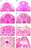
Figure 1
Figure 1
Hematoxylin and eosin staining of sectioned control and SB fetuses at various stages of gestation. (A,C,E,G) Cross-sections through the lumbar spinal area from E13.5 to P1 in control animals showing normal development of the spinal cord (dotted line), with laterally located dorsal root ganglia (open arrows), and surrounding skeletal and soft tissues. (B,D,F,H) Sections through the central part of age-matched litter mate fetuses/neonates with SB. At E13.5 (B), an intact dorsally-open spinal cord (dotted line) faces the amniotic cavity. The grey matter (purple), located dorsally, can be distinguished from the ventrally located white matter (light red). Dorsal root ganglia are located ventrally (open arrows) as a consequence of the failure of neural tube closure. At E16.5 (D), the dorsally-exposed spinal cord (dotted line) exhibits mild to moderate superficial neural tissue abrasion and hemorrhage, mostly located laterally (open arrowheads), probably resulting from contact with the uterine wall. At E18.5 (F), considerable loss of spinal cord tissue (dotted line) is visible, with only a few remnants of grey matter (purple) remaining. In contrast, more ventrally located white matter (light red) and dorsal root ganglia (open arrows) are mostly present. At birth (H), all exposed neural tissues have been lost and dorsal root ganglia are no longer discernible. The bare dorsal aspect of the vertebral body (open arrowheads) faces the amniotic cavity. Abbreviations: do, dorsal; vb, vertebral body; ve, ventral. Scale bars: 250 μm in A and B; 500 μm in C-H.
Figure 2
Figure 2
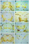
Neurofilament immunostaining of sectioned control and SB fetuses at various stages of gestation. (A,C,E,G) Cross-sections through the lumbar spinal area from E13.5 to P1 in control animals. The neurofilament staining method allows good visualization of sensory roots (s) projecting into the dorsal part of the spinal cord, motor roots (m) exiting the spinal cord ventrally, and dorsal root ganglia (open arrows) located ventro-laterally to the spinal cord. These structures characteristically exhibit a brownish coloration, whereas the spinal cord tissue stains light yellow. (B,D,F,H) Sections through the central part of age-matched litter mate fetuses/neonates with SB. At gestational stages E13.5, E15.5 and E17.5 (B,D,F), sensory roots, motor roots, and dorsal root ganglia (open arrows) are clearly visible and appear to be intact. As a consequence of failed neural tube closure, the sensory roots demonstrate an s-shaped kinking (particularly visible in D) and the dorsal root ganglia are located in a more ventral position than in controls. Location of motor roots is similar to that in normal controls. At birth (H), only remnants of neural tissue remain. Note the few neural bundles (probably motor root) that persist (arrowhead). Dorsal root ganglia (open arrows) are much smaller than in controls at this stage (G). Scale bars: 200 μm.
Figure 3
Figure 3
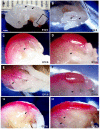
DiI tracer studies of control and SB fetuses at various stages of gestation. (A) Lateral and (B) dorsal/caudal views of a SB fetus (E12.5) immediately after insertion of DiI crystals (arrowheads). The lumbo-sacral area including the SB lesion and adjacent closed spinal cord (black box in A) is shown in top view in (B). DiI crystal within the MMC is indicated by left hand arrowhead in B. Two other crystals have been inserted into closed spinal cord rostral to the SB lesion. The dotted lines delineate the hindlimbs. A 20 g needle tip is shown for size comparison (top right in B). (C-H) Specimens photographed several weeks following insertion of DiI crystals. E12.5 fetuses (C,D) show a peripheral nerve exiting the spinal cord and running into the hindlimb (arrowheads) in both control (C) and SB (D) fetuses. E15.5 fetuses (E,F) show a very similar appearance as at E12.5, with a nerve entering the hindlimb in control and SB fetuses (arrowheads). At E17.5 (G,H), a nerve exiting the spinal cord and projecting into the hindlimb (arrowhead) is visible in the control fetus (G), whereas neural projections into the hindlimb (dotted line) from the SB lesion are not longer visible (H), although nerves can be seen exiting the spinal cord at levels rostral to the SB lesion (arrowheads in H). Scale bars: 1 mm in A, 0.7 mm in B, 0.35 mm in C and D, 0.7 mm in E, 0.55 mm in F, 0.65 mm in G and H.

Comment in

  • Fetal spina bifida.
    McLone DG. McLone DG. J Neurosurg. 2007 Mar;106(3 Suppl):211-2; discussion 212. doi: 10.3171/ped.2007.106.3.211. J Neurosurg. 2007. PMID: 17465387 No abstract available.

References

    1. Adzick NS, Harrison MR. Fetal surgical therapy. Lancet. 1994;343:897–902. - PubMed
    1. Asher JH, Jr., Harrison RW, Morell R, Carey ML, Friedman TB. Effects of Pax3 modifier genes on craniofacial morphology, pigmentation, and viability: A murine model of Waardenburg syndrome variation. Genomics. 1996;34:285–298. - PubMed
    1. Bruner JP, Tulipan N, Paschall RL, Boehm FH, Walsh WF, Silva SR, et al. Fetal surgery for myelomeningocele and the incidence of shunt- dependent hydrocephalus. JAMA. 1999;282:1819–1825. - PubMed
    1. Copp AJ, Greene NDE, Murdoch JN. The genetic basis of mammalian neurulation. Nat Rev Genet. 2003;4:784–793. - PubMed
    1. Correia-Pinto J, Reis JL, Hutchins GM, Baptista MJ, Estevao-Costa J, Flake AW, et al. In utero meconium exposure increases spinal cord necrosis in a rat model of myelomeningocele. J Pediatr Surg. 2002;37:488–492. - PubMed

Publication types