. 2007 Apr;3(4):e60.
doi: 10.1371/journal.ppat.0030060.

A novel TLR4-mediated signaling pathway leading to IL-6 responses in human bladder epithelial cells

Affiliations

A novel TLR4-mediated signaling pathway leading to IL-6 responses in human bladder epithelial cells

Jeongmin Song et al. PLoS Pathog. 2007 Apr.

Abstract

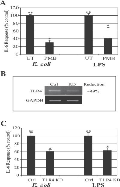
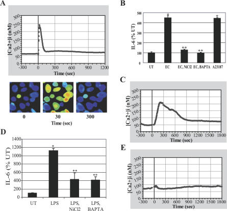
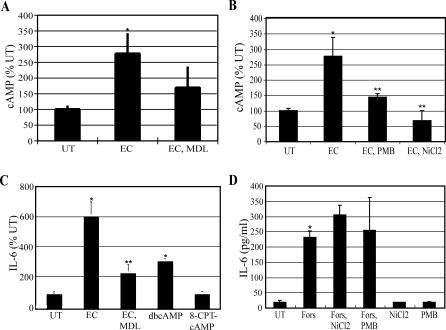
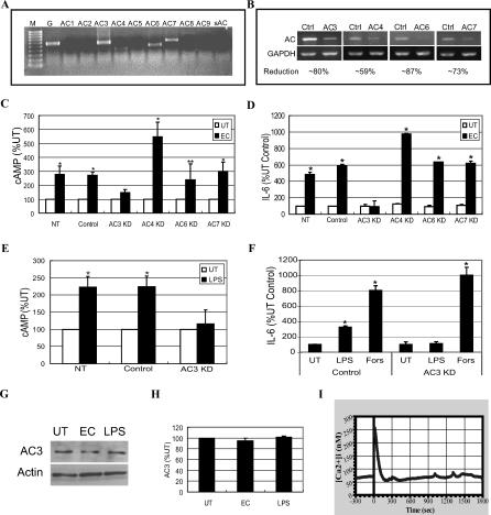
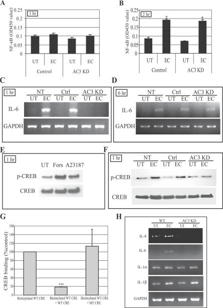
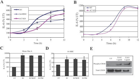
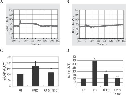
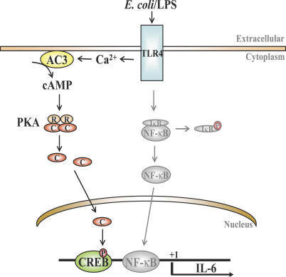
The vigorous cytokine response of immune cells to Gram-negative bacteria is primarily mediated by a recognition molecule, Toll-like receptor 4 (TLR4), which recognizes lipopolysaccharide (LPS) and initiates a series of intracellular NF-kappaB-associated signaling events. Recently, bladder epithelial cells (BECs) were reported to express TLR4 and to evoke a vigorous cytokine response upon exposure to LPS. We examined intracellular signaling events in human BECs leading to the production of IL-6, a major urinary cytokine, following activation by Escherichia coli and isolated LPS. We observed that in addition to the classical NF-kappaB-associated pathway, TLR4 triggers a distinct and more rapid signaling response involving, sequentially, Ca(2+), adenylyl cyclase 3-generated cAMP, and a transcriptional factor, cAMP response element-binding protein. This capacity of BECs to mobilize secondary messengers and evoke a more rapid IL-6 response might be critical in their role as first responders to microbial challenge in the urinary tract.


Conflict of interest statement

Competing interests. The authors have declared that no competing interests exist.

Figures

Figure 1
Figure 1. IL-6 Response of BECs to Type 1 Fimbriated E. coli Is Largely Elicited by LPS and Involves TLR4
(A) IL-6 secretion by BECs in response to E. coli (100 multiplicity of infection) or purified LPS (100 μg/ml). When specified, E. coli and purified LPS were pretreated with 1 μg/ml PMB for 30 min. **, p < 0.001 relative to values of untreated (UT) BECs; *, p < 0.03 relative to E. coli or LPS-treated BECs. (B) RT-PCR of control-transfected BECs (Ctrl) and TLR4 KD BECs. Glyseraldehyde-3-phosphate dehydrogenase (GAPDH) was employed as a loading control. (C) IL-6 secretion of control-transfected BECs (Ctrl) and TLR4 KD BECs after E. coli and LPS stimulation. **, p < 0.05 relative to UT control; *, p < 0.05 relative to E. coli-treated control or LPS control.
Figure 2
Figure 2. IL-6 Response of BECs to E. coli Is Preceded by an Increase in [Ca2+]i
(A) [Ca2+]i tracing of BECs before and after E. coli exposure. E. coli was added at time 0. (B) BEC IL-6 responses after E. coli exposure in the absence or presence of NiCl2 (2 mM) or BAPTA-AM (5 μM), or after calcium ionophore A23187 (1 μM) treatment without bacterial exposure. *, p < 0.001 relative to untreated (UT) BECs; **, p < 0.001 relative to E. coli (EC)–treated BECs. (C–E) BEC [Ca2+]i tracing before and after purified LPS treatment (C) or PMB pretreated LPS treatment (E). (D) BEC IL-6 responses following exposure to LPS in the absence or presence of NiCl2 or BAPTA-AM. *, p < 0.001 relative to UT BECs; **, p < 0.01 relative to LPS-treated BECs.
Figure 3
Figure 3. IL-6 Response of BECs to E. coli Is Associated with a Significant Increase in Intracellular cAMP Levels
(A and B) BEC cAMP production before and after E. coli exposure. When specified, BECs were pretreated with MDL-12,330A (MDL) (0.4 mM) or NiCl2 for 30 min, or E. coli was pretreated with PMB for 30 min. *p < 0.03 relative to untreated (UT) BECs; **p < 0.03 relative to E. coli (EC)–treated BECs. (C) IL-6 secretion by BECs was measured 6 h after exposure to E. coli, in the absence (EC) or presence of MDL-12,330A (EC, MDL), or after 6 h of treatment with 1 mM dibutyryl cAMP (dbcAMP), or 1 mM 8-(4-chloro-phenylthio)-2′-O-methyladenosine-3′-5′-cyclic monophosphate (8-CPT-cAMP) without bacterial exposure. *, p < 0.01 relative to UT; **, p < 0.02 relative to EC. (D) IL-6 secretion by BECs incubated for 6 h in the absence (UT) or presence of the AC-activator forskolin (Fors) (50 μM) with or without NiCl2 or PMB, or incubated with NiCl2 or PMB in the absence of forskolin. *, p < 0.01 relative to UT BECs.
Figure 4
Figure 4. AC-3 Is responsible for Mediating E. coli–Induced cAMP Production in BECs
(A) RT-PCR of BECs using primers specific for the ten known mammalian AC isoforms. Only AC-3, AC-4, AC-6, and AC-7 mRNA was expressed. GAPDH-specific RT-PCR (lane G) was used as a loading control. M, marker; sAC, soluble AC. (B) RT-PCR of control-transfected BECs (Ctrl) and AC-3, AC-4, AC-6, or AC-7 KD BECs. GAPDH-specific RT-PCR was used as a loading control. (C and D) Intracellular cAMP production (C) and IL-6 secretion (D) by non-transfected BECs (NT), control-transfected BECs (Control), or AC-3, AC-4, AC-6, or AC-7 KD BECs left untreated (UT) or treated with E. coli (EC). *, p < 0.005 and **, p < 0.02 relative to respective UT values. (E and F) Intracellular cAMP production (E) and IL-6 secretion (F) by non-transfected BECs (NT), control-transfected BECs (Control), or AC-3 KD BECs were measured in the absence (UT) or presence of E. coli LPS, or presence of forskolin (Fors) without LPS. *, p < 0.003 relative to respective UT values. (G) BEC AC-3–specific Western blot before (UT) and after (EC) E. coli exposure for 1 h, or exposure to E. coli LPS for 6 h. An actin-specific Western blot was used as a loading control. (H) Densitometric analysis of AC-3–specific Western blots, using ImageJ software. (I) [Ca2+]i tracing in AC-3 KD BECs before and after E. coli exposure. E. coli was added at time 0.
Figure 5
Figure 5. cAMP-Mediated Phosphorylation of CREB and Binding of CREB to CRE Oligonucleotides
(A and B) NF-κB nuclear translocation in control-transfected BECs and AC-3 KD BECs before and after E. coli (EC) exposure for 1 h (A) or 2 h (B). UT, untreated. (C and D) IL-6 message levels in non-transfected (NT), control-transfected (Ctrl), and AC-3 KD BECs before and after E. coli exposure for 1 h (C) or 6 h (D) as measured by RT-PCR. (E) Western blot of CREB phosphorylation levels (p-CREB) in UT BECs, and forskolin (Fors)– or calcium ionophore A23187 (A23187)–treated BECs. The treatment was for 1 h. (F) Western blot showing CREB phosphorylation of NT, Ctrl, and AC-3 KD BECs before and after 1 h E. coli exposure. (G) CREB binding to CRE site of the IL-6 promoter. Nuclear extracts of BECs exposed for 1 h to E. coli ORN103(pSH2) were incubated with biotinylated WT CRE oligonucleotides in the absence (Biotinylated WT CRE) or presence of specific (Biotinylated WT CRE + WT CRE) or non-specific (Biotinylated WT CRE + MT CRE) oligonucleotide competitors. ***, p < 0.0001. (H) Expression analysis of mRNA levels of various genes with CRE sites in their promoter. WT and AC-3 KD BECs were incubated for 1 h with E. coli ORN103(pSH2), and then total RNA was collected from untreated (UI) and bacteria-treated BECs (EC) and subjected to RT-PCR. GAPDH was used as a loading control.
Figure 6
Figure 6. The IL-6 Response to E. coli of BECs Is the Product of Two Separate Signaling Pathways
(A) IL-6 secretion response to E. coli ORN103(pSH2) by WT and AC-3 KD BECs in the absence or presence of pyrrolidine dithiocarbamate (PDTC). (B) IL-6 secretion response to UPEC CI5 by WT and AC-3 KD BECs. (C and D) IL-6 secretion responses by Mono Mac 6 cell line (C) and 16-HBE cell line (D). Cells were treated for 12 h with E. coli ORN103(pSH2) in the absence (EC) or presence of NiCl2 or PKA inhibitor (PKI). *, p < 0.03 relative to untreated (UT) value. Identical results were obtained after 6 h of treatment with E. coli ORN103(pSH2). (E) Increased CREB phosphorylation in BECs in response to TLR2 and TLR3 ligands. Western blotting for phospho-CREB and total-CREB in UT BECs or BECs treated with either a TLR2 ligand (lipoteichoic acid) or a TLR3 ligand (polyinosine-polycytidylic acid).
Figure 7
Figure 7. The IL-6 Response of Primary Human BECs to UPEC Is Linked to [Ca2+]i and cAMP Increase
(A) [Ca2+]i tracing before and after exposure of primary human BECs to UPEC strain CI5. (B) [Ca2+]i tracing of the primary human BECs pretreated with NiCl2 for 30 min. UPEC was added at time 0. (C and D) Intracellular cAMP production (C), or IL-6 secretion (D) by the primary human BECs left untreated (UT) or treated with E. coli CI5 (UPEC) in the absence or presence of NiCl2 (UPEC, NiCl2), or treated with E. coli ORN103 (EC). *, p < 0.03 relative to UT value. **, p < 0.03 relative to UPEC (CI5)–treated value.
Figure 8
Figure 8. Proposed Model for TLR4 Signaling in BECs
The proposed rapidly induced second messenger– and CREB-mediated pathway (dark line) as well as the classical NF-κB (grey line) are shown. Both pathways are triggered by TLR4. P, phosphorylation; R and C, regulatory and catalytic subunits of PKA.

Similar articles

Cited by

References

    1. Akira S, Uematsu S, Takeuchi O. Pathogen recognition and innate immunity. Cell. 2006;124:783–801. - PubMed
    1. Kawai T, Akira S. TLR signaling. Cell Death Differ. 2006;13:816–825. - PubMed
    1. Schilling JD, Martin SM, Hunstad DA, Patel KP, Mulvey MA, et al. CD14- and Toll-like receptor-dependent activation of bladder epithelial cells by lipopolysaccharide and type 1 piliated Escherichia coli . Infect Immun. 2003;71:1470–1480. - PMC - PubMed
    1. Agace W, Hedges S, Andersson U, Andersson J, Ceska M, et al. Selective cytokine production by epithelial cells following exposure to Escherichia coli . Infect Immun. 1993;61:602–609. - PMC - PubMed
    1. Haraoka M, Hang L, Frendeus B, Godaly G, Burdick M, et al. Neutrophil recruitment and resistance to urinary tract infection. J Infect Dis. 1999;180:1220–1229. - PubMed

Publication types

MeSH terms