. 2007 Apr 27:7:38.
doi: 10.1186/1471-213X-7-38.

The lin-35/Rb and RNAi pathways cooperate to regulate a key cell cycle transition in C. elegans

Affiliations

The lin-35/Rb and RNAi pathways cooperate to regulate a key cell cycle transition in C. elegans

Jimmy Ouellet et al. BMC Dev Biol. .

Abstract

Background: The Retinoblastoma gene product (Rb) has been shown to regulate the transcription of key genes involved in cell growth and proliferation. Consistent with this, mutations in Rb are associated with numerous types of cancer making it a critical tumour suppressor gene. Its function is conferred through a large multiprotein complex that exhibits a dual function in both activation and repression of gene targets. In C. elegans, the Rb orthologue lin-35 functions redundantly with other transcriptional regulators to appropriately specify both vulval and pharyngeal cell fates.

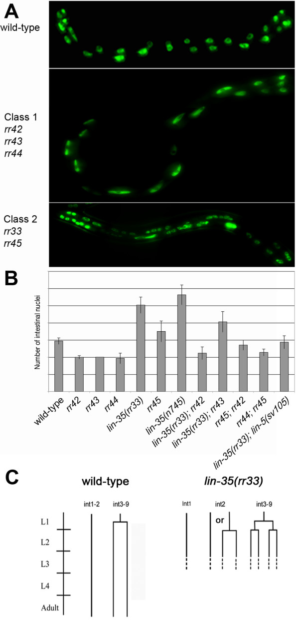
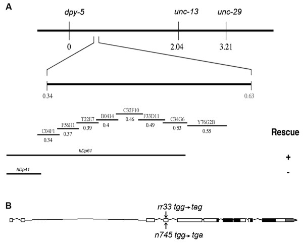
Results: In C. elegans the intestinal cells must alter their cell cycle from the mitotic cell divisions typical of embryogenesis to karyokinesis and then endoreplication, which facilitates growth during larval development. While screening for genes that affect the ability of the intestinal cells to appropriately make this cell cycle transition during post-embryonic development, we isolated mutants that either compromise this switch and remain mononucleate, or cause these cells to undergo multiple rounds of nuclear division. Among these mutants we identified a novel allele of lin-35/Rb, while we also found that the components of the synMuv B complex, which are involved in vulval specification, are also required to properly regulate the developmentally-controlled cell cycle transition typical of these intestinal cells during larval development. More importantly, our work uncovered a role for certain members of the pathways involved in RNAi in mediating the efficient transition between these cell cycle programs, suggesting that lin-35/Rb cooperates with these RNAi components. Furthermore, our findings suggest that met-2, a methyltransferase as well as hpl-1 and hpl-2, two C. elegans homologues of the heterochromatin protein HP1 are also required for this transition.

Conclusion: Our findings are consistent with lin-35/Rb, synMuv and RNAi components cooperating, probably through their additive effects on chromatin modification, to appropriately modulate the expression of genes that are required to switch from the karyokinesis cell cycle to endoreplication; a highly specified growth pathway in the intestinal epithelium. The lin-35/Rb repressor complex may be required to initiate this process, while components of the RNAi machinery positively reinforce this repression.


Figures

Figure 1
Figure 1
Two classes of mutants with altered numbers of intestinal nuclei. (A) Shows a wild-type (upper panel), a class 1 mutant with fewer than the wild-type number of intestinal nuclei (rr44) (middle) and class 2 mutant with extra intestinal nuclei (lin-35(rr33)) (lower panel). All animals were at the L4 larval stage and express the intestinal-specific elt-2::GFP. Anterior is left in all pictures. (B) Genetic interaction between the class 1 and class 2 mutants. All mutants with less intestinal nuclei (class 1) are epistatic to the mutants with supernumerary nuclear divisions (class 2). The double mutant lin-35(rr33); rr45 is synthetic lethal, therefore it is not represented on this graph. (C) Lineage analysis of the intestinal cells in lin-35(rr33) mutants. The diagram is representative of observations collected from 8 independent larvae from the initiation of post-embryonic development up to the end of the L2 stage. Horizontal lines represent intestinal nuclear divisions, while the vertical lines represent time.
Figure 2
Figure 2
Mapping and cloning of the rr33 mutant. (A) SNP-SNIP mapping as well as three-factor mapping approaches were used to map rr33 to the center of linkage group I, close to dpy-5. This region contains several well-defined free-duplications of which hDp61 rescued the rr33 mutant (+) whereas hDp41 did not (-). C32F10.2(RNAi) phenocopies the rr33 mutant phenotype. (B) A schematic representation of the C32F10.2 lin-35/Rb gene with black boxes representing the predicted pocket binding domains and the gray box indicating the 3' UTR. The location and the precise nucleotide changes in rr33 and n745 allele are indicated by arrows.
Figure 3
Figure 3
Inactivation of components in the synMuv B gene class enhances the lin-35(rr33) intestinal defect. (A) lin-35(rr33) animals were fed with bacterial clones that corresponded to the various components of the synMuv B complex and the number of intestinal nuclei were scored in L4 larvae 48 hrs later. The nhr-2 gene encodes a nuclear hormone receptor that has not been implicated in the synMuv B complex and was used as a negative control. (B) Feeding RNAi of some of the synMuv B genes was performed on wild-type animals and they also cause an slight increase in intestinal nuclei number, although to a lesser extent than lin-35(rr33) alone. The asterisk denotes a Student t-Test value of < 0.05 compared to lin-35(rr33) in A and wild-type in B. (C) A representative example of the multinucleate intestinal cells found in double mutant lin-35(rr33); dpl-1(RNAi) and lin-35(rr33); efl-1(RNAi) animals. The arrowheads indicate the intestinal cell boundaries. (D) The number of multinucleate cells was monitored in various genetic backgrounds. In wild type, all the intestinal cells of the Int3-9 rings have either 1 or 2 intestinal nuclei.
Figure 4
Figure 4
lin-35 mutants affect cyclin expression and intestinal nuclear ploidy. (A) qRT-PCR was performed on wild-type and lin-35(rr33) L1 larvae and the levels of cyclin expression were determined compared to wild-type. The bars represent 4 independent qRT-PCR reactions from RNA isolated from wild type and lin-35(rr33) animals. A positive value indicates an increase in gene expression compared to wild-type, while a negative value indicates a relative decrease in gene expression. The standard deviation is derived from the 4 independent trials. (B) DNA quantification was performed on the intestinal nuclei of both mutant and wild-type young adult hermaphrodites stained with propidium iodide. Each diamond represents a lin-35(r33) mutant intestinal nucleus and each square represents an individual wild type intestinal nucleus.
Figure 5
Figure 5
Components of the RNAi pathways cooperate with lin-35/Rb to regulate intestinal nuclear divisions. (A) lin-35(rr33) L4 animals were fed with bacterial clones that corresponded to the various components of the RNAi pathways and the number of intestinal nuclei was scored in L4 larvae of the next generation. (B) RNAi of some PTGS components was also performed on wild-type worms. The asterisk denotes a Student t-Test value of < 0.05 compared to lin-35(rr33) in A and wild-type in B. (C) Examples of the multinucleate intestinal cells typically generated in the double mutants between lin-35(rr33) and various components of the RNAi pathways. Some cells can exceed 12 nuclei/per cell. The arrowheads indicate the intestinal cell boundaries. (D) The number of multinucleate cells was monitored in various genetic backgrounds. In wild type, all the intestinal cells of the Int3-9 rings have either 1 or 2 intestinal nuclei.
Figure 6
Figure 6
Genetic regulation of cell cycle progression in the intestinal lineage of C. elegans. (A) All the animals depicted express an intestinal-specific GFP marker (elt-2::GFP), which is expressed from embryogenesis to the adult stage. The E blastomere is marked with a white asterisk as the transgene is not expressed at this early stage. The different types of intestinal cell cycle are marked under the schematic life cycle and the arrows indicate the developmental stage during which they occur. Black rectangles represent each molt. Anterior is up in all the figures and all the images are shown at the same magnification to represent the growth of the animal. (B) Proposed model for the lin-35/synMuv B complex in the regulation of the cell cycle transitions typical of the intestinal cell lineage during the L1 stage. See the text for details.

References

    1. Blagosklonny MV, Pardee AB. The restriction point of the cell cycle. Cell Cycle. 2002;1:103–110. - PubMed
    1. Lee WH, Bookstein R, Hong F, Young LJ, Shew JY, Lee EY. Human retinoblastoma susceptibility gene: cloning, identification, and sequence. Science. 1987;235:1394–1399. doi: 10.1126/science.3823889. - DOI - PubMed
    1. Giacinti C, Giordano A. RB and cell cycle progression. Oncogene. 2006;25:5220–5227. doi: 10.1038/sj.onc.1209615. - DOI - PubMed
    1. Ach RA, Durfee T, Miller AB, Taranto P, Hanley-Bowdoin L, Zambryski PC, Gruissem W. RRB1 and RRB2 encode maize retinoblastoma-related proteins that interact with a plant D-type cyclin and geminivirus replication protein. Mol Cell Biol. 1997;17:5077–5086. - PMC - PubMed
    1. Du W, Vidal M, Xie JE, Dyson N. RBF, a novel RB-related gene that regulates E2F activity and interacts with cyclin E in Drosophila. Genes Dev. 1996;10:1206–1218. - PubMed

Publication types

MeSH terms