Review
. 2008 Feb 4:1192:134-50.
doi: 10.1016/j.brainres.2007.03.044. Epub 2007 Mar 20.

Have we achieved a unified model of photoreceptor cell fate specification in vertebrates?

Affiliations
Review

Have we achieved a unified model of photoreceptor cell fate specification in vertebrates?

Ruben Adler et al. Brain Res. .

Abstract

How does a retinal progenitor choose to differentiate as a rod or a cone and, if it becomes a cone, which one of their different subtypes? The mechanisms of photoreceptor cell fate specification and differentiation have been extensively investigated in a variety of animal model systems, including human and non-human primates, rodents (mice and rats), chickens, frogs (Xenopus) and fish. It appears timely to discuss whether it is possible to synthesize the resulting information into a unified model applicable to all vertebrates. In this review we focus on several widely used experimental animal model systems to highlight differences in photoreceptor properties among species, the diversity of developmental strategies and solutions that vertebrates use to create retinas with photoreceptors that are adapted to the visual needs of their species, and the limitations of the methods currently available for the investigation of photoreceptor cell fate specification. Based on these considerations, we conclude that we are not yet ready to construct a unified model of photoreceptor cell fate specification in the developing vertebrate retina.


Figures

Figure 1
Figure 1
Hypothetical models of lineage relationships between rods (RH1 opsin) and cones expressing LWS opsin (red), RH2 opsin (green), SWS2 opsin (blue) and SWS1 opsin (violet). The homogeneity of the outer nuclear layer (ONL) in the early embryo would be consistent with the existence of an initial population of uncommitted photoreceptor progenitors (A). Heterogeneity associated with the segregation of rod and cone lineages could emerge at late stages of photoreceptor differentiation (A, a1), or during the long interval between photoreceptor generation and their advanced (“terminal”) differentiation (A, a2, a3). The heterogeneity of the early ONL would be consistent with a model that postulates early segregation of rods and cones (B), or even of rods and each separate cone subtype (C).
Figure 2
Figure 2
Molecular phylogeny of vertebrate photoreceptor opsin genes. Beginning with an ancestral opsin gene approximately 700 million years ago, a series of four gene duplication events with subsequent diversification produced five opsin gene families with spectral sensitivities ranging from longest (red) to shortest (ultraviolet) wavelengths: LWS/MWS, RH1, RH2, SWS2, and SWS1. The position of the branch point along the horizontal axis from left to right indicates the relative evolutionary age, from oldest to youngest, respectively. The most closely related are RH1 (rhodopsin) and RH2, which diverged approximately 500 million years ago. The LWS/MWS family is the oldest and most divergent of the five opsin gene families.
Figure 3
Figure 3
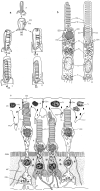
Morphology of cone and rod photoreceptors. The development [a] and structure [b] of a “typical” vertebrate cone (C) and rod (R) photoreceptor. [a] Development of the outer segment. The visual pigment is in membranous discs of the outer segment, which derive from infoldings of the plasma membrane of a microtubule-based, ciliated appendage. In rods, the discs pinch off completely from the plasma membrane, but in cones the discs are open to the extracellular environment. [b] Differentiated rod and cone photoreceptors. Note that the synaptic terminals of cones and rods are usually more distinct than in this illustration, in that cone pedicles are larger with multiple ribbon synapses and rod spherules are smaller with a single ribbon synapse. Abbreviations: F, flagellum (more accurately, cilium); FM, folds of the flagellar (i.e., ciliary) membrane; BB, basal body; PD, presumptive disc; OS, outer segment; D, disc; OD, oil droplet; Dn, dendrite (more accurately, calycal process); EM, ellipsoid mitochondria; IS, inner segment; P, paraboloid; N, nucleus; S, synapses. (c) The lamprey retina offers examples of ‘hybrid’ photoreceptor types. All photoreceptors have cone-like morphology, with open outer segment discs and synaptic terminals with the morphology typical of cone pedicles. However, the shorter type of photoreceptor has rod-like properties (rhodopsin visual pigment and scotopic sensitivity and kinetics). Abbreviations: PC, pigment cells; Dm, desmosome; Ds, disc stratification; PCP, pigment cell process; OBM, outer boundary membrane (outer limiting membrane); MF, Müller fiber; OSL, outer synaptic layer. Reprinted with permission from Vinnikov, Y.A. 1982. Evolution of Receptor Cells. Cytological, Membranous and Molecular Levels. (Translated from Russian by Nicholas Bobrov). Molecular Biology, Biochemistry and Biophysics, vol. 34, Springer-Verlag, Berlin Heidelberg New York, 141 pp.

Similar articles

Cited by

References

    1. Adler R. A model of retinal cell differentiation in the chick embryo. Prog Retinal Eye Res. 2000;20:529–557. - PubMed
    1. Adler R. Challenges in the study of neuronal differentiation: A view from the embryonic eye. Dev Dyn. 2005 - PMC - PubMed
    1. Ahmad I, Acharya HR, Rogers JA, Shibata A, Smithgall TE, Dooley CM. The role of NeuroD as a differentiation factor in the mammalian retina. J Mol Neurosci. 1998;11:165–178. - PubMed
    1. Ahnelt PK, Kolb H. The mammalian photoreceptor mosaic-adaptive design. Prog Retin Eye Res. 2000;19:711–777. - PubMed
    1. Allison WT, Dann SG, Helvik JV, Bradley C, Moyer HD, Hawryshyn CW. Ontogeny of ultraviolet-sensitive cones in the retina of rainbow trout (Oncorhynchus mykiss) J Comp Neurol. 2003;461:294–306. - PubMed