

Chondrocyte moves: clever strategies?
- PMID: 17467303
- PMCID: PMC1994785
- DOI: 10.1016/j.joca.2007.02.022
Chondrocyte moves: clever strategies?
Abstract
Goals: To review the literature on chondrocyte movements and to develop plausible hypothesis for further work.
Design: Chondrocyte movements are herein defined as translocations of the cell body. A brief overview of cell migration in other cell types is presented to set the stage for a discussion of chondrocyte moves; this includes a discussion of the challenges that cells find when moving within tissues. Reports of isolated chondrocyte migration in vitro (isolated cell systems) and ex vivo (cartilage organ cultures) are then summarized, followed by a discussion of recent studies that infer chondrocyte movements in vivo.
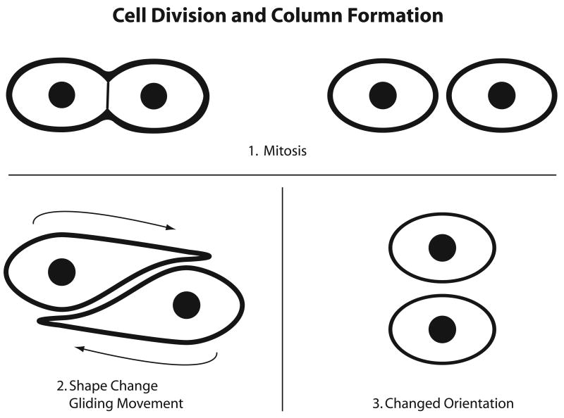
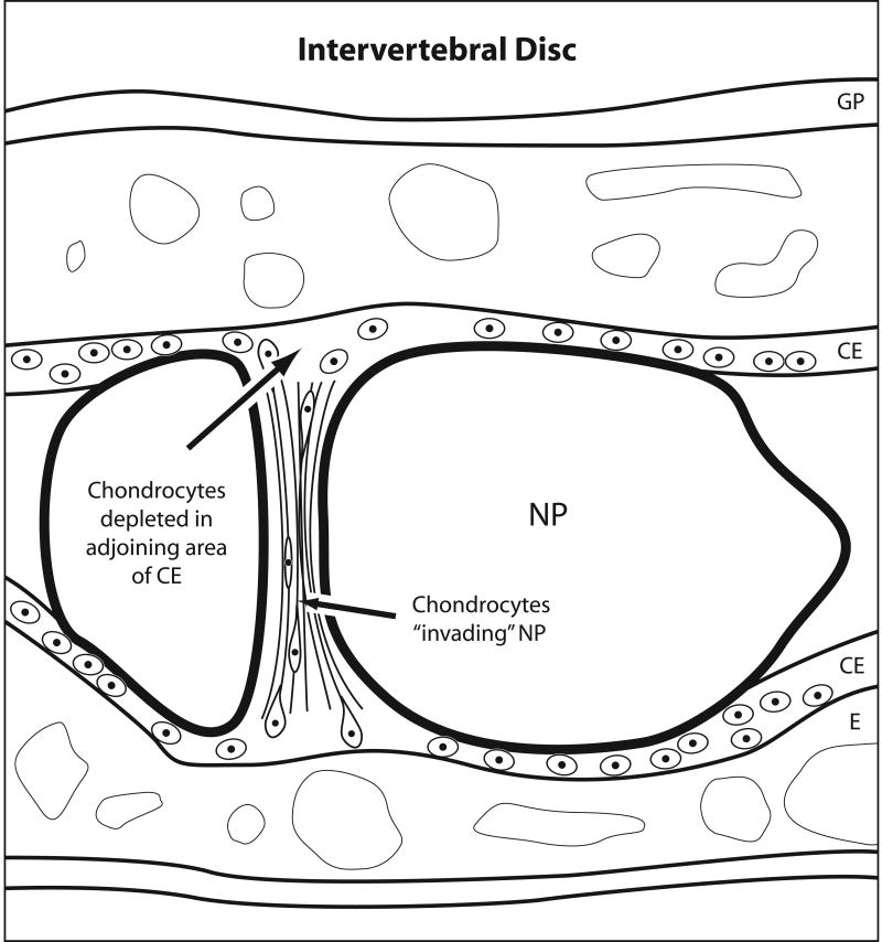
Results: Investigators from different laboratories have observed chondrocyte motility in vitro. I became interested in the question of whether articular chondrocytes retained their phenotype during their migratory excursions. We devised a simple method to separate migratory and stationary chondrocytes and then showed that migratory chondrocytes synthesized collagen II but not I--consistent with a differentiated phenotype. Our time-lapse video microscopy studies showed that the cells displayed appropriate movement kinetics, albeit with low speed and directionality. Similarly, others have presented data consistent with slow movement of chondrocytes out of cartilage explants. It is important to decipher whether these in vitro movements reflect physiological states and if so, which events are simulated. Examples of in vivo studies that have inferred chondrocyte movements include those describing rotational or gliding movements of chondrocytes in the proliferative zone of the growth plate and its importance in the growth process; and the notion that chondrocytes move from the cartilage endplates to the nucleus pulposus (NP) in the spine of rabbits and rats during development. Such studies are consistent with the hypothesis that chondrocytes exhibit highly controlled and specialized movements during tissue growth and remodeling in vivo. On the other hand, the cartilage explant studies elicit interest in the possibility that matrix injuries resulting in disruption of the collagen network of adult cartilages provide a permissive environment for chondrocyte motility.
Conclusions: The case for in vivo chondrocyte motility remains to be proven. However, the in vitro and in vivo data on chondrocyte movements present an argument for further thought and studies in this area.
Figures

References
-
- Lauffenburger DA, Horowitz AF. Cell Migration: A Physically Integrated Molecular Process. Cell. 1996;34:359–69. - PubMed
-
- Stossel TP. On the Crawling of Animal Cells. Science. 1993;260:1086–94. - PubMed
-
- Ridley AJ, Schwartz MA, Burridge K, Firtel RA, Ginsberg MH, et al. Cell Migration: Integrating signals from front to back. Science. 2003;302:1704–09. - PubMed
-
- Vicente-Manzanares M, Webb DJ, Horwitz AR. Cell migration at a glance. J Cell Science. 2005;118:4917–19. - PubMed
-
- Clemmons DR, Maile LA. Minireview: Integral membrane proteins that function coordinately with the insulin – like growth factor I receptor to regulate intracellular signaling. Endocrinology. 2003;144:1664–70. - PubMed
Publication types
MeSH terms
Grants and funding
LinkOut - more resources
Full Text Sources
Other Literature Sources
Miscellaneous
