. 2007 Jun 15;306(2):493-504.
doi: 10.1016/j.ydbio.2007.03.027. Epub 2007 Mar 27.

Cellular mechanisms of Müllerian duct formation in the mouse

Affiliations

Cellular mechanisms of Müllerian duct formation in the mouse

Grant D Orvis et al. Dev Biol. .

Abstract

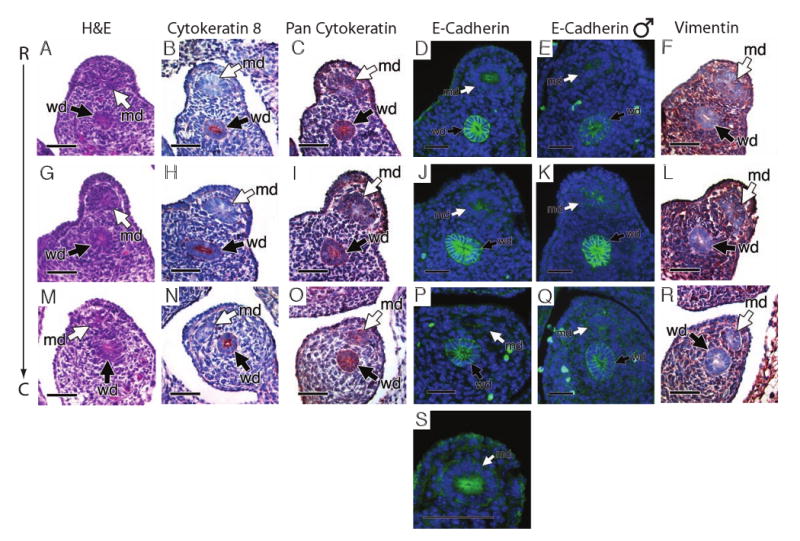
Regardless of their sex chromosome karyotype, amniotes develop two pairs of genital ducts, the Wolffian and Müllerian ducts. As the Müllerian duct forms, its growing tip is intimately associated with the Wolffian duct as it elongates to the urogenital sinus. Previous studies have shown that the presence of the Wolffian duct is required for the development and maintenance of the Müllerian duct. The Müllerian duct is known to form by invagination of the coelomic epithelium, but the mechanism for its elongation to the urogenital sinus remains to be defined. Using genetic fate mapping, we demonstrate that the Wolffian duct does not contribute cells to the Müllerian duct. Experimental embryological manipulations and molecular studies show that precursor cells at the caudal tip of the Müllerian duct proliferate to deposit a cord of cells along the length of the urogenital ridge. Furthermore, immunohistochemical analysis reveals that the cells of the developing Müllerian duct are mesoepithelial when deposited, and subsequently differentiate into an epithelial tube and eventually the female reproductive tract. Our studies define cellular and molecular mechanisms for Müllerian duct formation.


Figures

Fig. 1
Fig. 1
Staging Müllerian duct development. Lim1lz/+ expression in the Wolffian duct and growing Müllerian duct of females (A, C, E, G) and males (B, D, F, H). Whole mount lateral view (A and B) and ventral view (C-H) of Lim1lz/+ urogenital ridges. Müllerian duct precursor cells arise from the coelomic epithelium at tail somite 19 (TS19) in both females (A) and males (B). By TS22, both females (C) and males (D) have accomplished Müllerian duct invagination from the coelomic epithelium and contacted the Wolffian duct. The Müllerian duct has completed half of its elongation to the urogenital sinus at TS28 (E, F) and reached the urogenital sinus by TS34 (G, H). k, kidney (metanephros); md, Müllerian duct; mt, mesonephric tubules; o, ovary; t, testis; wd, Wolffian duct. Scale bar: 200 μM in A-D; 500 μM in E-H.
Fig. 2
Fig. 2
Analysis of growing Müllerian duct in relation to Wolffian duct at E13.0 in a male embryo. Whole mount urogenital ridge of a Lim1lz/+ embryo (A). (B-F) Cross-section of age matched wild-type embryo stained with hematoxylin and eosin. In the most rostral region, mesenchymal cells that form a sworl pattern surround the Müllerian duct (B). The Müllerian duct is located lateral in relation to the Wolffian duct (B). At E13.0, the Müllerian duct has crossed over the Wolffian duct to become located medially in relation to the Wolffian duct (C, D, E). (C, D) The Müllerian duct is separated from the Wolffian duct by mesenchymal cells, but these mesenchymal cells do not yet form a sworl around the Müllerian duct. Just rostral of the growing tip, the Müllerian duct is separated from the Wolffian duct (E). At the growing tip, cells of the Müllerian duct are in physical contact with cells of the Wolffian duct (F). g, gonad; md, Müllerian duct; wd, Wolffian duct. Scale bar: 20 μM.
Fig. 3
Fig. 3
Immunohistochemical analysis at E12.5 in wild-type tissue. The Müllerian duct was analyzed at three separate regions; rostral to the growing tip (A, B, C, D, E), at the growing tip (F, G, H, I, J) and caudal to the tip (K, L, M, N, O). (A, F, K) Hematoxylin and eosin staining. Cytokeratin 8, a marker of epithelium, was detected in the Wolffian duct, but not the developing Müllerian duct (B, G, L) as well as E-cadherin (D, I, N). The Wolffian duct also expressed pan-cytokeratin and the Müllerian duct showed weak expression (C, H, M). The Müllerian duct expresses vimentin, a marker of mesenchyme, at both its caudal tip and rostral region while the Wolffian duct does not express vimentin (E, J, O). High magnification of the vimentin stained tip reveals high expression of vimentin in the cells of the Müllerian duct tip (P). c, caudal; md, Müllerian duct; r, rostral; wd, Wolffian duct. Scale bar: 20 μM in A-O, 10μM in P.
Fig. 4
Fig. 4
Immunohistochemical analysis of XX (A-D, F-J, L-P, R, S) and XY (E, K, Q) embryos at E13.5 in wild-type tissue. The Müllerian duct was analyzed at three separate regions, rostral (A, B, C, D, E, F), middle (G, H, I, J, K, L) and caudal (M, N, O, P, Q, R). (A, G, M) Hematoxylin and eosin staining. The Wolffian duct, but not Müllerian duct expresses the epithelial marker cytokeratin 8 (B, H, N). Expression of pan-cytokeratin can be detected in the Wolffian duct and weakly in the Müllerian duct (C, I, O). The Müllerian duct weakly expresses the mesenchymal marker, vimentin, which is not expressed by the Wolffian duct (F, L, R). The Wolffian duct also expresses the epithelial cell marker E-cadherin in both males and females (D, E, J, K, P, Q). High magnification in a female embryo (S) reveals the most rostral portion of the Müllerian duct is expressing E-cadherin in its apical region, which is not seen in the male (E). c, caudal; md, Müllerian duct; r, rostral; wd, Wolffian duct. Scale bar: 20 μM in A-R, 10μM in S.
Fig. 5
Fig. 5
Wolffian duct specific Cre reporter analysis. lacZ staining of Hoxb7-Cre; R26R mice (A-F). The entire length of the Wolffian duct expresses lacZ from E11.5 to E13.5. Whole mount urogenital ridge at E11.5 (A), E12.5 (C) and E13.5 (E). Cross-section of the urogenital ridge at E12.5 (B, D, F). At both its growing tip and rostral of the growing tip, the Müllerian duct does not express lacZ (B, D). md, Müllerian duct; wd, Wolffian duct. Scale bar: 20 μM.
Fig. 6
Fig. 6
Urogenital ridge recombinant explant culture and cell proliferation analysis. Whole mount urogenital ridge three days after culture and stained with X-gal (A, B, C, D). (A) Unmanipulated Lim1lz/+ control. (B) Negative recombinant in which both Müllerian ducts failed to grow into the wild-type caudal ridge. (C, D) Successful explant culture in which one (C) or both (D) Müllerian ducts grew past the point of manipulation into the wild-type caudal ridge. (E, F) Cross-section of successful recombinant explant culture. (E) Lim1lz/+ rostral ridge with both the Wolffian and Müllerian ducts expressing lacZ. (F) Wild-type caudal ridge with a Lim1lz/+ Müllerian duct, but wild-type, lacZ negative, Wolffian duct. (G-J) Cell proliferation analysis of the growing Müllerian duct tip at E12.5. Positive immunostaining for pH3 (G) and BrdU (H) in the rostral duct and pH3 (I) and BrdU (J) at the growing tip. E12.5 starting control for the Müllerian duct tip formation assay (K). The rostral Müllerian duct cells were removed from the proliferating tip cells and the ridge cultured. The isolated tip cells were able to form the Müllerian duct independent of the rostral duct cells (L). Arrowheads denote the point at which the Lim1lz/+ rostral ridge was cut. The light blue staining in the wild-type caudal ridges of B-D is background trapped in the tubules of the metanephric kidney. The line in K indicates the point at which the urogenital ridge was cut. g, gonad; k, kidney; md, Müllerian duct; wd, Wolffian duct. Scale bar: 20 μM.
Fig. 7
Fig. 7
A three phase model for Müllerian duct development. In the first phase, cells of the coelomic epithelium are specified to become Müllerian duct cells (A, B). After specification the second phase begins and these cells invaginate caudally towards the Wolffian duct (C, D). Once the Müllerian duct comes into contact with the Wolffian duct, the third phase begins (E, F) and the Müllerian duct elongates caudally, following the path of the Wolffian duct, towards the urogenital sinus. Blue cells; mesoepithelial Müllerian duct cells, red cells; proliferating Müllerian duct precursor cells, brown cells; coelomic epithelial cells, yellow cells; Wolffian epithelial cells. ce; coelomic epithelium, md; Müllerian duct, wd, Wolffian duct

References

    1. Carroll TJ, Park JS, Hayashi S, Majumdar A, McMahon AP. Wnt9b plays a central role in the regulation of mesenchymal to epithelial transitions underlying organogenesis of the mammalian urogenital system. Dev Cell. 2005;9:283–92. - PubMed
    1. Colas JF, S GC. Towards a cellular and molecular understanding of neurulation. Dev Dyn. 2001;221:117–145. - PubMed
    1. Dohr G, Tarmann T, Schiechl H. Different antigen expression on Wolffian and Mullerian cells in rat embryos as detected by monoclonal antibodies. Anat Embryol (Berl) 1987;176:239–42. - PubMed
    1. Drews U. Direct and mediated effects of testosterone: analysis of sex reversed mosaic mice heterozygous for testicular feminization. Cytogenet Cell Genet. 1998;80:68–74. - PubMed
    1. Dyche WJ. A comparative study of the differentiation and involution of the Mullerian duct and Wolffian duct in the male and female fetal mouse. J Morphol. 1979;162:175–209. - PubMed

Publication types