. 2007 Jul;189(13):4944-52.
doi: 10.1128/JB.01518-06. Epub 2007 Apr 27.

Specific bonds between an iron oxide surface and outer membrane cytochromes MtrC and OmcA from Shewanella oneidensis MR-1

Affiliations

Specific bonds between an iron oxide surface and outer membrane cytochromes MtrC and OmcA from Shewanella oneidensis MR-1

Brian H Lower et al. J Bacteriol. 2007 Jul.

Abstract

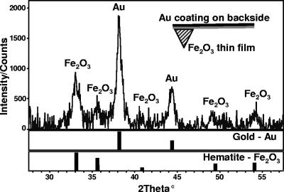
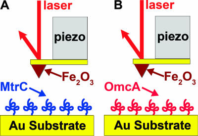
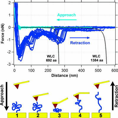
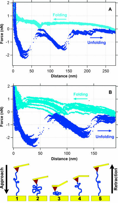
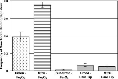
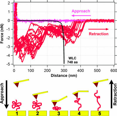
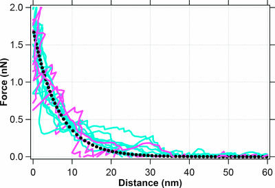
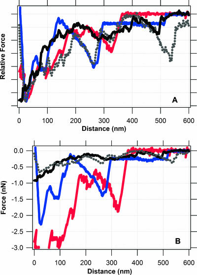
Shewanella oneidensis MR-1 is purported to express outer membrane cytochromes (e.g., MtrC and OmcA) that transfer electrons directly to Fe(III) in a mineral during anaerobic respiration. A prerequisite for this type of reaction would be the formation of a stable bond between a cytochrome and an iron oxide surface. Atomic force microscopy (AFM) was used to detect whether a specific bond forms between a hematite (Fe(2)O(3)) thin film, created with oxygen plasma-assisted molecular beam epitaxy, and recombinant MtrC or OmcA molecules coupled to gold substrates. Force spectra displayed a unique force signature indicative of a specific bond between each cytochrome and the hematite surface. The strength of the OmcA-hematite bond was approximately twice that of the MtrC-hematite bond, but direct binding to hematite was twice as favorable for MtrC. Reversible folding/unfolding reactions were observed for mechanically denatured MtrC molecules bound to hematite. The force measurements for the hematite-cytochrome pairs were compared to spectra collected for an iron oxide and S. oneidensis under anaerobic conditions. There is a strong correlation between the whole-cell and pure-protein force spectra, suggesting that the unique binding attributes of each cytochrome complement one another and allow both MtrC and OmcA to play a prominent role in the transfer of electrons to Fe(III) in minerals. Finally, by comparing the magnitudes of binding force for the whole-cell versus pure-protein data, we were able to estimate that a single bacterium of S. oneidensis (2 by 0.5 microm) expresses approximately 10(4) cytochromes on its outer surface.


Figures

FIG. 1.
FIG. 1.
X-ray diffraction pattern of an AFM cantilever that was coated with a hematite thin film using oxygen plasma-assisted molecular beam epitaxy. Gold (Au) and hematite (Fe2O3) references are shown for comparison. The Au signal originates from a coating applied to the “backside” (see schematic inset) of the cantilever by the manufacturer. The Au coating increases laser reflectivity for the AFM's optical lever detection, but it does not interfere with measurements of the force between the protein and hematite.
FIG. 2.
FIG. 2.
Schematic describing the experimental setup used to examine the interactions between hematite (Fe2O3) and purified S. oneidensis MR-1 outer membrane cytochromes MtrC (A) and OmcA (B). MtrC and OmcA are drawn arbitrarily here since their three-dimensional structures are not known. Purified recombinant proteins were covalently attached to a gold substrate via terminal Cys residues and were probed with a hematite-functionalized AFM tip in PBS buffer at pH 7.4.
FIG. 3.
FIG. 3.
Measurements of force between hematite and MtrC. The top graph shows 14 approach (light blue) and 14 retraction (dark blue) force curves. The dotted black lines correspond to the theoretical force-extension relationship for polypeptides composed of 692 (single MtrC molecule) and 1,384 (putative MtrC dimer) amino acids as predicted by the WLC model (equation 2). The bottom schematic illustrates one approach/retraction cycle, starting at frame 1 and ending at frame 5.
FIG. 4.
FIG. 4.
Folding and unfolding trajectories observed for MtrC during approach (light blue) and retraction (dark blue) cycles. Here, the hematite-coated tip was not fully retracted as described for Fig. 3, but rather only to a distance of 275 nm (A) or 200 nm (B). Under these conditions, the MtrC molecule does not detach from the tip but rather maintains a physical bond with the hematite-functionalized tip (depicted in the bottom schematic), allowing the observation of unfolding and refolding pathways of the polypeptide.
FIG. 5.
FIG. 5.
Plot showing the frequency of observing a binding event (i.e., sawtooth force signature) between S. oneidensis MR-1 cytochromes and hematite (Fe2O3). Shown are the average values (±standard errors) for OmcA-functionalized substrates and hematite-coated tips (n = 816), MtrC-functionalized substrates and hematite-coated tips (n = 881), bare substrates that were not coated with cytochromes and hematite-coated tips (n = 487), OmcA-functionalized substrates and bare Si3N4 tips (n = 468), and MtrC-functionalized substrates and bare Si3N4 tips (n = 633). These values include only those force curves for which the tip was completely separated from the polypeptide during an approach-retraction cycle, and they do not include folding-refolding cycles like those shown in Fig. 4.
FIG. 6.
FIG. 6.
Measurements of the force between hematite and OmcA. The top graph shows 14 approach (light red) and 14 retraction (dark red) force curves. The dotted black line corresponds to the theoretical force-extension relationship for a polypeptide composed of 748 amino acids (single OmcA molecule), as predicted by the WLC model (equation 2). The bottom schematic illustrates one approach/retraction cycle, starting at frame 1 and ending at frame 5.
FIG. 7.
FIG. 7.
Force curves for hematite approaching a MtrC-functionalized substrate (light blue) or an OmcA-functionalized substrate (light red). The dotted black curve is the theoretical steric force (equation 1) between a hematite-coated tip (60-nm radius) and a surface coated with a protein (thickness = 40 nm; density = 2.3 × 10¹⁶ molecules m−2).
FIG. 8.
FIG. 8.
Spectra for force-distance relationship between an Fe(III) oxide and either a purified cytochrome or S. oneidensis MR-1. Traces correspond to retraction curves for an Fe(III) oxide and each of the following: S. oneidensis MR-1 grown with Fe(III) as the terminal electron acceptor (black and gray curves), recombinant MtrC (blue curve), and recombinant OmcA (red curve). Curves for the purified cytochromes come from this study. The solid black curve was taken from reference , and the dotted gray curve was taken from reference . (A) Relative force-distance spectra where the y axis has been normalized such that the maximum force in each retraction curve is set to a value of 1. This procedure does not alter the distance data; it simply plots force on a relative scale. (B) Unaltered versions of the force spectra presented in panel A for comparison.

References

    1. Arnold, R. G., T. J. DiChristina, and M. R. Hoffmann. 1988. Reductive dissolution of Fe(III) oxides by Pseudomonas species 200. Biotechnol. Bioeng. 32:1081-1096. - PubMed
    1. Baumann, C. G., S. B. Smith, V. A. Bloomfield, and C. Bustamante. 1997. Ionic effects on the elasticity of single DNA molecules. Proc. Natl. Acad. Sci. USA 94:6185-6190. - PMC - PubMed
    1. Beliaev, A. S., D. A. Saffarini, J. L. McLaughlin, and D. Hunnicutt. 2001. MtrC, an outer membrane decahaem c cytochrome required for metal reduction in Shewanella putrefaciens MR-1. Mol. Microbiol. 39:722-730. - PubMed
    1. Bell, G. 1978. Models for the specific adhesion of cells to cells. Science 200:618-627. - PubMed
    1. Bemis, J. E., B. B. Akhremitchev, and G. C. Walker. 1999. Single polymer chain elongation by atomic force microscopy. Langmuir 15:2799-2805.

Publication types

LinkOut - more resources