. 2007 Aug 1;110(3):1036-42.
doi: 10.1182/blood-2007-02-076919. Epub 2007 Apr 27.

The ring-infected erythrocyte surface antigen (RESA) of Plasmodium falciparum stabilizes spectrin tetramers and suppresses further invasion

Affiliations

The ring-infected erythrocyte surface antigen (RESA) of Plasmodium falciparum stabilizes spectrin tetramers and suppresses further invasion

Xinhong Pei et al. Blood. .

Abstract

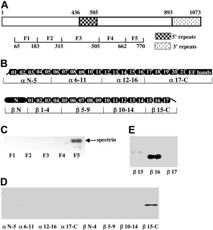
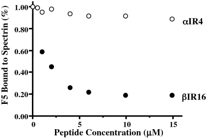
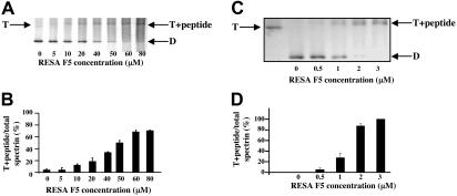
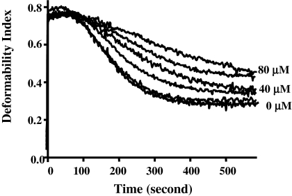
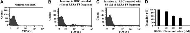
The malaria parasite Plasmodium falciparum releases the ring-infected erythrocyte surface antigen (RESA) inside the red cell on entry. The protein migrates to the host cell membrane, where it binds to spectrin, but neither the nature of the interaction nor its functional consequences have previously been defined. Here, we identify the binding motifs involved in the interaction and describe a possible function. We have found that spectrin binds to a 108-amino acid fragment (residues 663-770) of RESA, and that this RESA fragment binds to repeat 16 of the beta-chain, close to the labile dimer-dimer self-association site. We further show that the RESA fragment stabilizes the spectrin tetramer against dissociation into its constituent dimers, both in situ and in solution. This is accompanied by enhanced resistance of the cell to both mechanical and thermal degradation. Resealed erythrocytes containing RESA(663-770) display resistance to invasion by merozoites of P falciparum. We infer that the evolutionary advantage of RESA to the parasite lies in its ability to prevent invasion of cells that are already host to a developing parasite, as well as possibly to guard the cell against thermal damage at the elevated body temperatures prevailing in febrile crises.


Figures

Figure 1
Figure 1
Identification of spectrin-RESA binding sites. (A) Schematic representation of RESA protein and recombinant fragments. The locations of the 5′ and 3′ repeat regions, and the positions in the sequence of the expressed fragments are indicated. (B) Schematic representation of the spectrin α- and β-chains, showing the domain structure, and locations in the sequence of expressed recombinant fragments. The boundaries of all spectrin fragments and single repeats were defined by SMART annotations. (C) Binding of spectrin dimer to RESA fragments. Spectrin dimer was incubated for 30 minutes at room temperature with each of the MBP-tagged RESA fragments, and binding was assessed by pull-down assay; the bound fragment was detected by blotting with antispectrin antibody after SDS-PAGE. (D) Binding of recombinant spectrin fragments to the RESA F5 fragment. Recombinant GST-tagged spectrin fragments were incubated with RESA F5 and binding was assayed as described for panel C, using anti-GST antibody for detection. (E) Binding of single GST-tagged β-spectrin repeats to RESA F5 fragment. Binding assays were performed as above. Note binding to βR16 only.
Figure 2
Figure 2
Inhibition of binding of RESA F5 fragment to spectrin dimer by spectrin repeat βR16, assessed by ELISA. RESA F5 was incubated with increasing concentrations of βR16 or αR4 before addition to immobilized spectrin dimer in the ELISA wells. RESA F5 binding to spectrin is inhibited by βR16, but not by αR4.
Figure 3
Figure 3
Stabilization of spectrin tetramer by RESA F5 fragment. Panels A and B show the composition of spectrin extracted at 37°C from erythrocyte membranes previously resealed without or with the indicated concentrations of RESA F5. Spectrin components were separated by nondenaturing gel electrophoresis in the cold. The positions of migration of dimer (D), tetramer (T), and the tetramer-RESA-F5 complex (T + peptide) are indicated. Panels C and D show suppression of conversion of spectrin tetramer to dimer by RESA F5 in solution. Spectrin tetramer in the low-ionic-strength extraction medium was incubated with the RESA fragment at the indicated concentrations at 37°C for 1 hour, followed by gel electrophoresis, as before. The best-fit of binding data in situ at 37°C shown in panel B corresponds to an association constant of approximately 2 × 10⁴ M−1, while that of binding data shown in panel D results in an association constant of approximately 1 × 10⁶ M−1. (B,D) Results represent the mean value of triplicate samples.
Figure 4
Figure 4
Effect of intracellular RESA F5 fragment on red cell membrane stability. RESA F5 at concentrations of 0, 5, 20, 40, 60, and 80 μM was added to ghosts before resealing. Membrane mechanical stability of resealed ghosts was measured by ektacytometry. The deformability index (DI) measures the deformation of the membranes under constant shear stress. The decay of DI with time is due to progressive breakdown of the cells to vesicles, and thus reflects the extent of shear resistance. The fragmentation profiles for 0, 40, and 80 μM are labeled. It should be noted there is a progression in membrane stability with increasing concentrations of RESA peptide.
Figure 5
Figure 5
Effect of membrane-bound RESA F5 fragment on malarial invasion. The proportion of cells infected by P falciparum in culture was assayed by flow cytometry, using YOYO-1 to stain parasite DNA. (A) Uninfected ghosts. (B) Control ghosts (no RESA fragment) exposed to parasites in culture. (C) Ghosts containing 80 μM RESA F5. (D) Dependence of invasion efficiency on intracellular RESA F5 concentration. At the highest concentration of RESA F5 used (80 μM), at which invasion is suppressed by about 60%, the calculated occupancy of spectrin sites in the cell by the peptide is about 60%. Results represent the mean value of triplicate samples.

References

    1. Cooke BM, Mohandas N, Coppel RL. The malaria-infected red blood cell: structural and functional changes. Adv Parasitol. 2001;50:1–86. - PMC - PubMed
    1. Hiller NL, Bhattacharjee S, van Ooij C, et al. A host-targeting signal in virulence proteins reveals a secretome in malarial infection. Science. 2004;306:1934–1937. - PubMed
    1. Marti M, Baum J, Rug M, Tilley L, Cowman AF. Signal-mediated export of proteins from the malaria parasite to the host erythrocyte. J Cell Biol. 2005;171:587–592. - PMC - PubMed
    1. Sargeant TJ, Marti M, Caler E, et al. Lineage-specific expansion of proteins exported to erythrocytes in malaria parasites. Genome Biol. 2006;7:R12. - PMC - PubMed
    1. Baruch DI, Pasloske BL, Singh HB, et al. Cloning the P. falciparum gene encoding PfEMP1, a malarial variant antigen and adherence receptor on the surface of parasitized human erythrocytes. Cell. 1995;82:77–87. - PubMed

Publication types

MeSH terms

Substances