

Incidence and visual outcome of endophthalmitis associated with intraocular foreign bodies
- PMID: 17468878
- PMCID: PMC2206251
- DOI: 10.1007/s00417-007-0586-5
Incidence and visual outcome of endophthalmitis associated with intraocular foreign bodies
Abstract
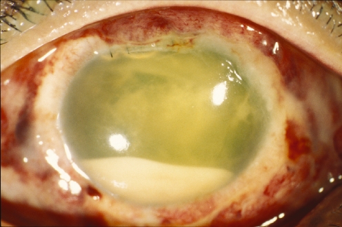
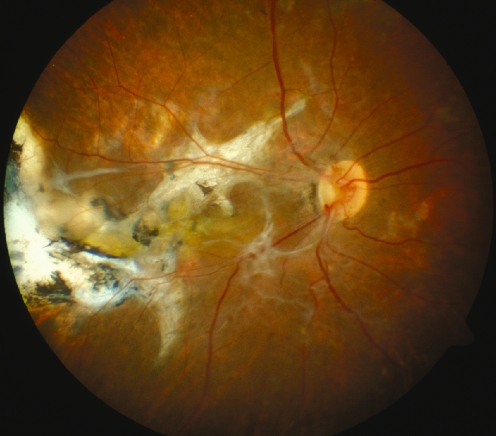
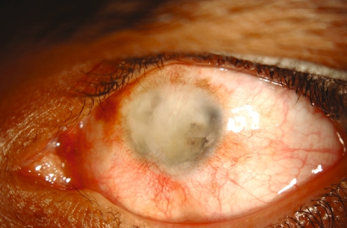
Purpose: To determine the risk factors and visual outcome of endophthalmitis associated with traumatic intraocular foreign body (IOFB) removal and its allied management.
Methods: A retrospective review was conducted of patients with penetrating eye trauma and retained IOFB with associated endophthalmitis managed at King Khaled Eye Specialist Hospital over a 22 year period (1983 to 2004).
Results: There were 589 eyes of 565 patients (90.3% male; 9.7% female) which sustained ocular trauma and had retained IOFB that required management. Forty-four eyes (7.5%) developed clinical evidence of endophthalmitis at some point after trauma. From these 44 eyes, initial presenting visual acuity (VA) of 20/200 or better was recorded in 8 eyes (18.1%) and the remaining 36 eyes (81.9%) had VA ranging from 20/400 to light perception. Eleven eyes (25%) underwent IOFB removal and repair within 24 hours after trauma while 33 eyes (75%) had similar procedures done 24 hours or more after trauma. Thirty-one eyes (70%) underwent primary pars plana vitrectomy (PPV) at the time of removal of posteriorly located IOFBs. Definite positive cultures were obtained from 17 eyes (38.6%). Over a mean follow-up of 24.8 months, 21 eyes (47.7%) had improved VA, 6 eyes (13.6%) maintained presenting VA while 17 eyes (38.7%) had deterioration of their VA, including 10 eyes (22.7%) that were left with no light perception (NLP) vision. After the treatment of endophthalmitis, 20 eyes (45.4%) had VA of 20/200 or better at their last follow-up. Four eyes (12.9%) from the vitrectomy group (31 eyes) and 5 eyes (45.4%) from non-vitrectomy (11 eyes) group had final VA of NLP. Predictive factors for the good visual outcome included good initial presenting VA, early surgical intervention to remove IOFB (within 24 hours), and PPV. Predictors of poor visual outcome included IOFB removal 48 hours or later, posterior location and no PPV for the posteriorly located IOFB.
Conclusions: Delayed removal of IOFB following trauma may result in a significant increase in the development of clinical endophthalmitis. Other risk factors for poor visual outcome may include poor initial presenting VA, posterior location of IOFB and no vitrectomy at the time of IOFB removal.
Figures
References
-
- {'text': '', 'ref_index': 1, 'ids': [{'type': 'PubMed', 'value': '8414406', 'is_inner': True, 'url': 'https://pubmed.ncbi.nlm.nih.gov/8414406/'}]}
- Thompson JT, Parver LM, Enger CL et al (1993) Infectious endophthalmitis after penetrating injuries with retained intraocular foreign bodies. National Eye Trauma System. Ophthalmology 100:1468–1474 - PubMed
-
- {'text': '', 'ref_index': 1, 'ids': [{'type': 'DOI', 'value': '10.1046/j.1442-9071.2004.00759.x', 'is_inner': False, 'url': 'https://doi.org/10.1046/j.1442-9071.2004.00759.x'}, {'type': 'PubMed', 'value': '14746594', 'is_inner': True, 'url': 'https://pubmed.ncbi.nlm.nih.gov/14746594/'}]}
- Azad RV, Kumar N, Sharma YR, Vohra R (2004) Role of prophylactic scleral buckling in the management of retained intraocular foreign bodies. Clin Exp Ophthalmol 32:58–61 - PubMed
-
- {'text': '', 'ref_index': 1, 'ids': [{'type': 'PubMed', 'value': '3262852', 'is_inner': True, 'url': 'https://pubmed.ncbi.nlm.nih.gov/3262852/'}]}
- Williams DF, Mieler WF, Abrams GW, Lewis H (1988) Results and prognostic factors in penetrating ocular injuries with retained intraocular foreign bodies. Ophthalmology 90:1318–1322 - PubMed
-
- {'text': '', 'ref_index': 1, 'ids': [{'type': 'PubMed', 'value': '11192838', 'is_inner': True, 'url': 'https://pubmed.ncbi.nlm.nih.gov/11192838/'}]}
- El-Asrar AM, Al-Amro SA, Khan NM, Kangave D (2000) Visual outcome and prognostic factors after vitrectomy for posterior segment foreign bodies. Eur J Ophthalmol 10:304–311 - PubMed
-
- {'text': '', 'ref_index': 1, 'ids': [{'type': 'PubMed', 'value': '2710504', 'is_inner': True, 'url': 'https://pubmed.ncbi.nlm.nih.gov/2710504/'}]}
- Behrens-Baumann W, Praetorius G (1989) Intraocular foreign bodies: 297 consecutive cases. Ophthalmologica 198:84–88 - PubMed
MeSH terms
LinkOut - more resources
Full Text Sources
Miscellaneous
