Review
. 2007 Aug;454(5):759-76.
doi: 10.1007/s00424-007-0247-x. Epub 2007 Apr 28.

Signal transduction and information processing in mammalian taste buds

Affiliations
Review

Signal transduction and information processing in mammalian taste buds

Stephen D Roper. Pflugers Arch. 2007 Aug.

Abstract

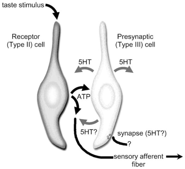
The molecular machinery for chemosensory transduction in taste buds has received considerable attention within the last decade. Consequently, we now know a great deal about sweet, bitter, and umami taste mechanisms and are gaining ground rapidly on salty and sour transduction. Sweet, bitter, and umami tastes are transduced by G-protein-coupled receptors. Salty taste may be transduced by epithelial Na channels similar to those found in renal tissues. Sour transduction appears to be initiated by intracellular acidification acting on acid-sensitive membrane proteins. Once a taste signal is generated in a taste cell, the subsequent steps involve secretion of neurotransmitters, including ATP and serotonin. It is now recognized that the cells responding to sweet, bitter, and umami taste stimuli do not possess synapses and instead secrete the neurotransmitter ATP via a novel mechanism not involving conventional vesicular exocytosis. ATP is believed to excite primary sensory afferent fibers that convey gustatory signals to the brain. In contrast, taste cells that do have synapses release serotonin in response to gustatory stimulation. The postsynaptic targets of serotonin have not yet been identified. Finally, ATP secreted from receptor cells also acts on neighboring taste cells to stimulate their release of serotonin. This suggests that there is important information processing and signal coding taking place in the mammalian taste bud after gustatory stimulation.


Figures

Fig. 1
Fig. 1
Calculations discussed in Johanningsmeier et al. [12]
Fig. 2
Fig. 2
Schematic drawing showing a taste cell stimulated with acetic acid. Acetic acid in solution dissociates into a mixture of protonated acetic acid (HAcetate), acetate, and protons (H+), the latter of which are bound to one or more water molecules (H3O+). HAcetate, being uncharged, penetrates the lipid plasma membrane and enters the cytosol. Inside the cell, HAcetate dissociates and releases H+ (which binds to H2O, H3O+) and acetate, acidifying the cytosol (lowering pHi)
Fig. 3
Fig. 3
Schematic drawing of the sweet GPCR dimer T1R2+T1R3, showing the multiple ligand binding sites. T1R2 is shown on the left, T1R3 is on the right. Details of the interactions, if any, between the extensive N termini of T1R2 and T1R3 are not known. The N termini possess one or more binding pocket(s) for saccharin, sucrose, and other sugars. Another ligand-binding pocket, located in T1R3 near the first transmembrane region, exists for certain sweet-tasting proteins such as brazzein. A third ligand pocket, embedded in the transmembrane (TM) regions of T1R3, is comprised of portions of TM3, 5, and 6. This site binds the artificial sweetener, cyclamate, but also is the binding site for antagonists such as saccharin (which at high concentrations is a sweet receptor antagonist) and lactisole
Fig. 4
Fig. 4
Taste stimulation for G protein-coupled taste receptors (e.g. sweet, bitter, umami) initiates two parallel streams of intracellular events. Several details are known for the PLCβ2-signaling stream (right). Considerably less is understood about the role of cyclic nucleotides in taste transduction (left), especially taste-evoked decreases in cAMP. The final events shown, depolarization leading to transmitter release, remain somewhat speculative
Fig. 5
Fig. 5
Cell–cell communication within the mammalian taste bud. Taste receptor (type II) cells (left) secrete ATP in response to gustatory stimulation. ATP acts on sensory afferent fibers as well as on adjacent presynaptic (type III) cells (right), causing the latter to release serotonin (5HT). Consequently, gustatory stimulation of taste buds elicits ATP and serotonin release. Presynaptic cells form synapses, possibly serotonergic, with targets that have not yet been identified with confidence. Postsynaptic sites for 5HT may include other taste cells (paracrine activation), sensory afferent fibers, or unidentified postsynaptic targets, as shown above
Fig. 6
Fig. 6
Schematic drawing of a mammalian taste bud illustrating hypothetical clustering of receptor (type II) and presynaptic (type III) cells for information coding. Left, nine representative taste cells (out of a total of 50 to 100, typical of taste buds) are depicted. A single sensory afferent axon is shown innervating a cluster of taste cells. For clarity, afferent innervation of other cells has been omitted. ATP secreted from receptor (type II) cells (light gray) excites afferent axons. ATP from receptor cells also stimulates presynaptic (type III) cells (dark gray). Middle, the cluster of cells contacting the afferent fiber is isolated for emphasis. Three receptor cells, each of which might express a similar taste GPCR, are shown for illustration. Activation of any of the receptor cells in the cluster would result in ATP secretion and excitation of the afferent fiber. ATP would also excite the presynaptic cell shown in the middle of the cluster. Right, a type I supporting cell ensheathing the cluster of taste cells is added. Type I cells express ecto-ATPase which would be expected to limit the diffusional spread of ATP throughout the taste bud, perhaps concentrating its actions on small clusters of cells as shown here. Even though each receptor cell expresses one type of taste GPCR, the information carried by the single sensory afferent (i.e., its “code”) might be ambiguous if not all the receptor cells in the cluster expressed identical taste GPCRs. Further, activity in the presynaptic (type III) cell would reflect stimulation of any of the nearby receptor cells. However, gustatory coding from the entire taste bud or multiple taste buds could be resolved if activity in numbers of sensory afferents were combined or averaged. Such a schema might explain how electrophysiological and functional imaging studies report the existence of taste cells that respond to multiple taste stimuli (here, presynaptic (type III) cells) alongside cells that respond only to one tastant (i.e., receptor (type II) cells). The schema would also explain the multiple taste sensitivity recorded from single afferent fibers

Similar articles

Cited by

References

    1. Simon SA, de Araujo I, Gutierrez R, Nicolelis MA. The neural mechanisms of gustation: a distributed processing code. Nat Rev Neurosci. 2006;7:890–901. - PubMed
    1. Meyerhof W. Elucidation of mammalian bitter taste. Rev Physiol Biochem Pharmacol. 2005;154:37–72. - PubMed
    1. Spector AC, Travers SP. The representation of taste quality in the mammalian nervous system. Behav Cogn Neurosci Rev. 2005;4:143–191. - PubMed
    1. Breslin PA, Huang L. Human taste: peripheral anatomy, taste transduction, and coding. Adv Oto-Rhino-Laryngol. 2006;63:152–190. - PubMed
    1. Gilbertson TA. Gustatory mechanisms for the detection of fat. Curr Opin Neurobiol. 1998;8:447–452. - PubMed

MeSH terms

LinkOut - more resources