

Up-regulation of p55 TNF alpha-receptor in dorsal root ganglia neurons following lumbar facet joint injury in rats
- PMID: 17468886
- PMCID: PMC2200776
- DOI: 10.1007/s00586-007-0365-3
Up-regulation of p55 TNF alpha-receptor in dorsal root ganglia neurons following lumbar facet joint injury in rats
Abstract
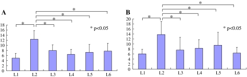
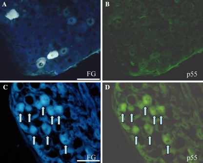
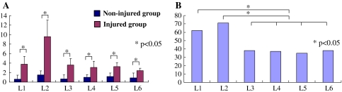
The rat L5/6 facet joint is multisegmentally innervated from the L1 to L6 dorsal root ganglia (DRG). Tumor necrosis factor (TNF) is a known mediator of inflammation. It has been reported that satellite cells are activated, produce TNF and surround DRG neurons innervating L5/6 facet joints after facet injury. In the current study, changes in TNF receptor (p55) expression in DRG neurons innervating the L5/6 facet joint following facet joint injury were investigated in rats using a retrograde neurotransport method followed by immunohistochemistry. Twenty rats were used for this study. Two crystals of Fluorogold (FG; neurotracer) were applied into the L5/6 facet joint. Seven days after surgery, the dorsal portion of the capsule was cut in the injured group (injured group n = 10). No injury was performed in the non-injured group (n = 10). Fourteen days after the first application of FG, bilateral DRGs from T13 to L6 levels were resected and sectioned. They were subsequently processed for p55 immunohistochemistry. The number of FG labeled neurons and number of FG labeled p55-immunoreactive (IR) neurons were counted. FG labeled DRG neurons innervating the L5/6 facet joint were distributed from ipsilateral L1 to L6 levels. Of FG labeled neurons, the ratio of DRG neurons immunoreactive for p55 in the injured group (50%) was significantly higher than that in the non-injured group (13%). The ratio of p55-IR neurons of FG labeled DRG neurons was significantly higher in total L1 and L2 DRGs than that in total L3, 4, 5 and 6 DRGs in the injured group (L1 and 2 DRG, 67%; L3, 4, 5 and 6 DRG, 37%, percentages of the total number of p55-IR neurons at L1 and L2 level or L3-6 level/the total number of FG-labeled neurons at L1 and L2 level or L3-6 level). These data suggest that up-regulation of p55 in DRG neurons may be involved in the sensory transmission from facet joint injury. Regulation of p55 in DRG neurons innervating the facet joint was different between upper DRG innervated via the paravertebral sympathetic trunks and lower DRG innervated via other direct routes.
Figures
Similar articles
-
Up-regulation of TNFalpha in DRG satellite cells following lumbar facet joint injury in rats.Eur Spine J. 2006 Jun;15(6):953-8. doi: 10.1007/s00586-005-1031-2. Epub 2006 Apr 29. Eur Spine J. 2006. PMID: 16758109 Free PMC article.
-
Phenotypic inflammation switch in rats shown by calcitonin gene-related peptide immunoreactive dorsal root ganglion neurons innervating the lumbar facet joints.Spine (Phila Pa 1976). 2001 May 1;26(9):1009-13. doi: 10.1097/00007632-200105010-00005. Spine (Phila Pa 1976). 2001. PMID: 11337618
-
Substance P and calcitonin gene-related peptide immunoreactive sensory DRG neurons innervating the lumbar facet joints in rats.Auton Neurosci. 2000 Dec 28;86(1-2):13-7. doi: 10.1016/S1566-0702(00)00194-6. Auton Neurosci. 2000. PMID: 11269919
-
Distribution and immunocytochemical characterization of dorsal root ganglion neurons innervating the lumbar intervertebral disc in rats: a review.Life Sci. 2004 Apr 9;74(21):2627-42. doi: 10.1016/j.lfs.2004.01.008. Life Sci. 2004. PMID: 15041445 Review.
-
Relationship of dorsal root ganglion to intervertebral foramen in lumbar region: an anatomical study and review of literature.J Neurosurg Sci. 2016 Sep;60(3):339-44. J Neurosurg Sci. 2016. PMID: 27402404 Review.
Cited by
-
Management of lumbar zygapophysial (facet) joint pain.World J Orthop. 2016 May 18;7(5):315-37. doi: 10.5312/wjo.v7.i5.315. eCollection 2016 May 18. World J Orthop. 2016. PMID: 27190760 Free PMC article.
-
Lumbar facet joint compressive injury induces lasting changes in local structure, nociceptive scores, and inflammatory mediators in a novel rat model.Pain Res Treat. 2012;2012:127636. doi: 10.1155/2012/127636. Epub 2012 Jun 28. Pain Res Treat. 2012. PMID: 22966427 Free PMC article.
-
Bilateral muscle fiber and nerve influences by TNF-alpha in response to unilateral muscle overuse - studies on TNF receptor expressions.BMC Musculoskelet Disord. 2017 Nov 28;18(1):498. doi: 10.1186/s12891-017-1796-6. BMC Musculoskelet Disord. 2017. PMID: 29183282 Free PMC article.
-
Enhanced BDNF and ROS in Mucosa of Lower Motor Neuron Lesioned Dog Bladder Following Somatic Motor Nerve Transfer.Cells. 2025 Mar 11;14(6):406. doi: 10.3390/cells14060406. Cells. 2025. PMID: 40136655 Free PMC article.
-
Reactive oxygen species mediate TNFR1 increase after TRPV1 activation in mouse DRG neurons.Mol Pain. 2009 Jun 17;5:31. doi: 10.1186/1744-8069-5-31. Mol Pain. 2009. PMID: 19531269 Free PMC article.
References
MeSH terms
Substances
LinkOut - more resources
Full Text Sources
