Multi-modal magnetic resonance imaging quantifies atherosclerosis and vascular dysfunction in patients with type 2 diabetes mellitus
- PMID: 17469043
- PMCID: PMC2243181
- DOI: 10.3132/dvdr.2007.005
Multi-modal magnetic resonance imaging quantifies atherosclerosis and vascular dysfunction in patients with type 2 diabetes mellitus
Abstract
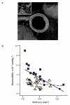
Vascular magnetic resonance imaging (MRI) is emerging as a powerful research tool. We studied 18 patients with type 2 diabetes mellitus and 20 controls (all with coronary artery disease). MRI measured distensibility, pulse wave velocity (PWV) and atherosclerosis in the aorta, and brachial artery flow-mediated dilatation (FMD). Patients with diabetes showed lower aortic distensibility (2.1 x 10(-3) vs . 3.5 x 10(-3) mmHg-1, p<0.01), faster PWV (8.8 vs ., 6.2 m/s, p<0.01) and impaired FMD (8.5% vs . 13.8%, p<0.05). Diabetes was an independent negative predictor of distensibility. Aortic atherosclerosis was similar in the two groups. There was a negative correlation between aortic distensibility and atherosclerosis in control subjects only, suggesting that other factors such as protein cross-linking may explain lower aortic distensibility in diabetes. MRI provides comprehensive vascular phenotyping in patients with type 2 diabetes and is likely to be useful in studies of disease progression and drug therapy.
Figures


References
-
- Zimmet P, Alberti KG, Shaw J. Global and societal implications of the diabetes epidemic. Nature. 2001;414:782–7. - PubMed
-
- Topol EJ, Nissen SE. Our preoccupation with coronary luminology. The dissociation between clinical and angiographic findings in ischemic heart disease. Circulation. 1995;92:2333–42. - PubMed
-
- Celermajer DS, Sorensen KE, Gooch VM, et al. Non-invasive detection of endothelial dysfunction in children and adults at risk of atherosclerosis. Lancet. 1992;340:1111–15. - PubMed
-
- Williams SB, Cusco JA, Roddy MA, Johnstone MT, Creager MA. Impaired nitric oxide-mediated vasodilation in patients with non-insulin-dependent diabetes mellitus. J Am Coll Cardiol. 1996;27:567–74. - PubMed
MeSH terms
Grants and funding
LinkOut - more resources
Full Text Sources
Medical

