. 2007 May;61(5):435-45.
doi: 10.1002/ana.21154.

TDP-43 immunoreactivity in hippocampal sclerosis and Alzheimer's disease

Affiliations

TDP-43 immunoreactivity in hippocampal sclerosis and Alzheimer's disease

Catalina Amador-Ortiz et al. Ann Neurol. 2007 May.

Abstract

Objective: This study aimed to determine the frequency of frontotemporal lobar degeneration with ubiquitinated inclusions (FTLD-U) in the setting of hippocampal sclerosis (HpScl) and Alzheimer's disease (AD) using immunohistochemistry for TAR DNA binding protein 43 (TDP-43), a putative marker for FTLD-U.

Methods: Initially, 21 cases of HpScl associated with a variety of other pathological processes and 74 cases of AD were screened for FTLD-U with TDP-43 immunohistochemistry. A confirmation study was performed on 93 additional AD cases. Specificity of TDP-43 antibodies was assessed using double-immunolabeling confocal microscopy, immunoelectron microscopy, and biochemistry.

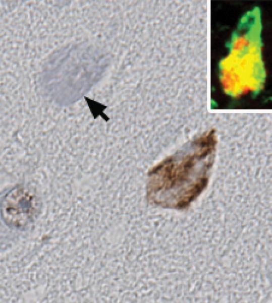
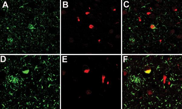
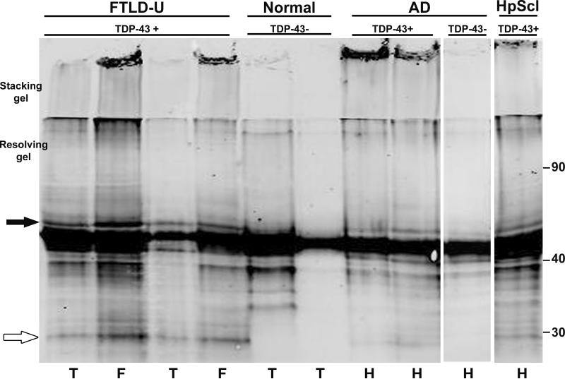
Results: TDP-43 immunoreactivity was detected in 71% of HpScl and 23% of AD cases. Double immunostaining of AD cases for TDP-43 and phospho-tau showed that the TDP-43-immunoreactive inclusions were usually distinct from neurofibrillary tangles. At the ultrastructural level, TDP-43 immunoreactivity in AD was associated with granular and filamentous cytosolic material and only occasionally associated with tau filaments. Western blots of AD cases showed a band that migrated at a higher molecular weight than normal TDP-43 that was not present in AD cases without TDP-43 immunoreactivity.

Interpretation: These results suggest that as many as 20% of AD cases and more than 70% of HpScl cases have pathology similar to that found in FTLD-U. Whether this represents concomitant FTLD-U or is analogous to colocalization of alpha-synuclein and tau in AD, reflecting a propensity for codeposition of abnormal protein conformers, remains to be determined.


Figures

Fig 1
Fig 1
Comparison of TDP-43 antibodies, (A) mouse monoclonal and (B) rabbit polyclonal, with immunohistochemistry of adjacent sections of the same case reveals neuronal cytoplasmic inclusions (inset at higher magnification) in a subset of neurons in the hippocampal dentate fascia. Note also the staining of normal nuclei and the absence of nuclear staining in neurons with cytoplasmic inclusions. Images are ×400 and inset ×800.
Fig 2
Fig 2
AD case with HpScl showing TDP-43 positive cytoplasmic inclusions and neurites in the dentate fascia (A), entorhinal cortex (B), nucleus accumbens (C), cingulate gyrus (D) and amygdala (E). Intranuclear neuronal inclusions are detected in the entorhinal cortex in some cases (F). Images are ×400 and inset ×800.
Fig 3
Fig 3
Distribution of TDP-43 immunoreactive inclusions. (A) The diffuse type has inclusions in the dentate fascia of the hippocampus (hp), the entorhinal cortex (erc), the occipitotemporal gyrus (otg) as well as the inferior temporal gyrus (itg). (B) The limbic type showed immunoreactivity limited to the dentate fascia and entorhinal cortex, with sparse or no involvement of the occipitotemporal gyrus.
Fig 4
Fig 4
In AD cases that did not have FTLD-U-like TDP-43 immunoreactivity, occasional NFTs in the subiculum (and less often the entorhinal cortex) have TDP-43 immunoreactivity. An adjacent extracellular NFT (arrow) shows no staining. Inset shows a confocal microscopic image of a NFT double stained for phosphotau (green) and TDP-43 (red) showing only partial overlap of phospho-tau and TDP-43 immunoreactivities in NFTs. Images are ×400
Fig 5
Fig 5
Double-labeling for phospho-tau (A and D) (green) and TDP-43 (B and E) (red) with merged images (C and F) shows two types of co-localization of phospho-tau and TDP-43. In Type 1 (D, E and F) there is overlap of the epitopes in some of the inclusions, while other inclusions are single labeled for either tau or TDP-43. In Type 2 (A, B and C) there is intermingling, but separation, of the two epitopes within the same neuron, as well as inclusions that are single labeled for either tau or TDP-43. All images are ×400.
Fig 6
Fig 6
Immunoelectron microscopy of TDP-43 shows a neuron (A) with an intranuclear inclusion (arrow), nucleolus (No) and cytoplasmic filaments part of a NFT (double arrows). L, lipofuscin. Bar, 1 μm. In (B), an enlargement of the boxed area in (A) shows that the intranuclear inclusion composed of granule coated filaments of 15−20-nm in diameter that are heavily labeled with TDP-43 (gold particles in circles). Chromatin at the nuclear membrane is unlabeled (arrow). Bar, 0.1 μm. In (C) enlargement of the NFT reveals paired helical filaments (arrows) and straight filaments (curved arrows) that are unlabeled for TDP-43. The nucleolus is also unlabeled (not shown). Bar, 0.15 μm. In (D) a neuronal with NFT has tau filaments in tightly packed bundles (arrows) and randomly oriented in the cytoplasm (*). L, lipofuscin. Bar, 1 μm. In (E) the boxed area in (D) is enlarged to show that the bundled filaments tightly packed with dense granular material and heavy labeling for TDP-43 (gold particles in circles), while the randomly oriented straight tau filaments are mostly unlabeled. Bar, 0.3 μm.
Fig 7
Fig 7
Western blot of urea fractions of hippocampal (H), temporal (T) and frontal cortices (F) from AD and HpScl TDP-43 immunoreactive cases (TDP-43+) exhibited abnormal bands of approximately 45 (black arrow) and 25 (white arrow) kDa, similar to what is seen in FTLD-U controls. This pattern was not found in AD and normal elderly individuals that did not have TDP-43 immunoreactive lesions (TDP-43−).

References

    1. McKhann GM, Albert MS, Grossman M, et al. Clinical and pathological diagnosis of frontotemporal dementia: report of the Work Group on Frontotemporal Dementia and Pick's Disease. Arch Neurol. 2001;58:1803–1809. - PubMed
    1. Bergmann M, Kuchelmeister K, Schmid KW, et al. Different variants of frontotemporal dementia: a neuropathological and immunohistochemical study. Acta Neuropathol (Berl) 1996;92:170–179. - PubMed
    1. Lipton AM, White CL, 3rd, Bigio EH. Frontotemporal lobar degeneration with motor neuron disease-type inclusions predominates in 76 cases of frontotemporal degeneration. Acta Neuropathol (Berl) 2004;108:379–385. - PubMed
    1. Mann DM, South PW, Snowden JS, Neary D. Dementia of frontal lobe type: neuropathology and immunohistochemistry. J Neurol Neurosurg Psychiatry. 1993;56:605–614. - PMC - PubMed
    1. Snowden JS, Neary D, Mann DM. Frontotemporal dementia. Br J Psychiatry. 2002;180:140–143. - PubMed

MeSH terms