Review
. 2007 May;133(3):535-56.
doi: 10.1037/0033-2909.133.3.535.

Social outcomes in childhood brain disorder: a heuristic integration of social neuroscience and developmental psychology

Affiliations
Review

Social outcomes in childhood brain disorder: a heuristic integration of social neuroscience and developmental psychology

Keith Owen Yeates et al. Psychol Bull. 2007 May.

Abstract

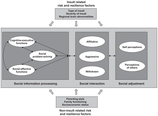
The authors propose a heuristic model of the social outcomes of childhood brain disorder that draws on models and methods from both the emerging field of social cognitive neuroscience and the study of social competence in developmental psychology/psychopathology. The heuristic model characterizes the relationships between social adjustment, peer interactions and relationships, social problem solving and communication, social-affective and cognitive-executive processes, and their neural substrates. The model is illustrated by research on a specific form of childhood brain disorder, traumatic brain injury. The heuristic model may promote research regarding the neural and cognitive-affective substrates of children's social development. It also may engender more precise methods of measuring impairments and disabilities in children with brain disorder and suggest ways to promote their social adaptation.


Figures

Figure 1
Figure 1
An integrative, heuristic model of social competence in children with brain disorder.
Figure 2
Figure 2
Brain regions implicated in social cognition and executive function. Panel a shows medial surface of the right hemisphere depicting the limbic lobe, laterobasal-cortical amygdala, orbital-medial frontal cortex, and hippocampus. The laterobasal-cortical amygdala and hippocampus are projected on the surface of the parahippocampal gyrus. Panel b shows lateral surface of right hemisphere depicting the superior temporal sulcus, superior temporal gyrus, fusiform gyrus, and temporal and frontal poles. Panel c shows midsaggital section depicting orbitofrontal cortex, medial prefrontal cortex, and anterior cingulate cortex. Adapted, with permission, from “Autism: A Window Onto the Development of the Social and the Analytic Brain,” by S. Baron-Cohen and M. K. Belmonte, 2005, Annual Review of Neuroscience, 28, p. 113. Copyright 2005 by Annual Reviews (www.annualreviews.org). Also adapted, with permission, from “The Limbic Lobe and Its Output Channels: Implications for Emotional Functions and Adaptive Behavior,” by L. Heimer and G. W. Van Hoesen, 2006, Neuroscience and Biobehavioral Reviews, 30, p. 135. Copyright 2006 by Elsevier.
Figure 3
Figure 3
Regional brain abnormalities following traumatic brain injury in children. Results are based on voxel-based morphometry analyses through use of statistical parametric mapping (SPM) techniques. Participants were 6 adolescents (mean age = 16 years, SD = 5.1), all with severe TBI (i.e., Glasgow Coma Scale scores of 8 or below) and associated frontal and temporal contusions, compared with 18 control subjects of similar age (3 control subjects per TBI patient within 2 years of age). The gray scale SPM plots at the top of the figure illustrate significant differences (p < .001) between TBI patients and controls. Darker shading represents areas with less density of gray matter pixels (i.e., greater atrophy) within a given voxel among TBI subjects as compared with controls. The red arrow points to where the most significant changes were observed. In the colored three-dimensional portrayal in the bottom portion of the figure, the areas in red represent the regions where significant differences were found in the density of gray matter pixels within the comparison voxels. Adapted, with permission, from “Structural Imaging” by E. D. Bigler, in Textbook of Traumatic Brain Injury (2nd ed.), 2005, Washington, DC: American Psychiatric Publishing. Copyright 2005 by American Psychiatric Publishing.
Figure 4
Figure 4
Proximity of basal ganglia, ventral striatum, and amygdala. Panel A depicts a coronal thin section (1 mm) 3 Tesla MRI (T1 inversion recovery sequence) at the level of the basal ganglia and amygdaloid complex also showing location of the superior temporal gyrus (STG; Panel D, bottom row, blue arrow). The highlighted region shown in the box is enlarged in Panel B and labeled according to Heimer’s (2003) classification system, as shown in Panel C. The coronal sections in the bottom two rows of Panel D are 1 mm apart, with the section in Panel A shown in the outline in the bottom row of Panel D (green oval). The blue arrow identifies the STG, which can be viewed in each subsequent coronal section, and the yellow arrow identifies the temporal stem. Red arrows show the position of the anterior commissure, and the green oval outlines the region of the ventral striatum, parahippocampal gyrus, and amygdala. Adapted, with permission, from “The Limbic Lobe and Its Output Channels: Implications for Emotional Functions and Adaptive Behavior,” by L. Heimer and G. W. Van Hoesen, 2006, Neuroscience and Biobehavioral Reviews, 30, p. 128. Copyright 2006 by Elsevier.

References

    1. Achenbach TM. Manual for the Child Behavior Checklis/4–18 and 1991 profile. Burlington, VT: University of Vermont, Department of Psychiatry; 1991.
    1. Adolphs R. The neurobiology of social cognition. Current Opinion in Neurobiology. 2001;11:231–239. - PubMed
    1. Adolphs R. Neural systems for recognizing emotion. Current Opinion in Neurobiology. 2002;12:169–177. - PubMed
    1. Adolphs R. Cognitive neuroscience of human social behaviour. Nature Reviews: Neuroscience. 2003;4:165–178. - PubMed
    1. Adolphs R, Baron-Cohen S, Tranel D. Impaired recognition of social emotions following amygdala damage. Journal of Cognitive Neuroscience. 2002;14:1264–1274. - PubMed

Publication types