Review
. 2007 Jun;8(4):373-99.
doi: 10.1016/j.sleep.2007.03.008. Epub 2007 Apr 30.

Clinical and neurobiological aspects of narcolepsy

Affiliations
Review

Clinical and neurobiological aspects of narcolepsy

Seiji Nishino. Sleep Med. 2007 Jun.

Abstract

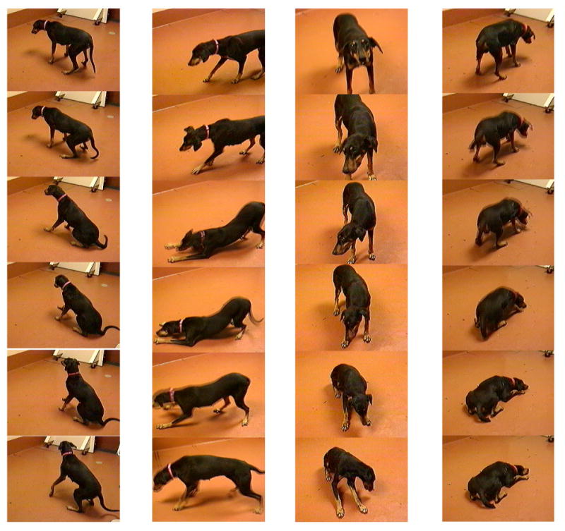
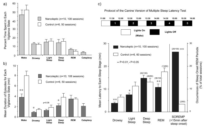
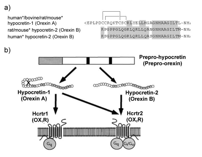
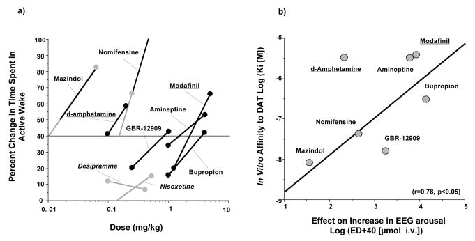
Narcolepsy is characterized by excessive daytime sleepiness (EDS), cataplexy and/or other dissociated manifestations of rapid eye movement (REM) sleep (hypnagogic hallucinations and sleep paralysis). Narcolepsy is currently treated with amphetamine-like central nervous system (CNS) stimulants (for EDS) and antidepressants (for cataplexy). Some other classes of compounds such as modafinil (a non-amphetamine wake-promoting compound for EDS) and gamma-hydroxybutyrate (GHB, a short-acting sedative for EDS/fragmented nighttime sleep and cataplexy) given at night are also employed. The major pathophysiology of human narcolepsy has been recently elucidated based on the discovery of narcolepsy genes in animals. Using forward (i.e., positional cloning in canine narcolepsy) and reverse (i.e., mouse gene knockout) genetics, the genes involved in the pathogenesis of narcolepsy (hypocretin/orexin ligand and its receptor) in animals have been identified. Hypocretins/orexins are novel hypothalamic neuropeptides also involved in various hypothalamic functions such as energy homeostasis and neuroendocrine functions. Mutations in hypocretin-related genes are rare in humans, but hypocretin-ligand deficiency is found in many narcolepsy-cataplexy cases. In this review, the clinical, pathophysiological and pharmacological aspects of narcolepsy are discussed.


Figures

Fig. 1
Fig. 1. Cataplectic attacks in Doberman pinschers
Emotional excitations, appetizing food or playing, easily elicit multiple cataplectic attacks in these animals. Most cataplexy attacks are bilateral (97.9%). Atonia initiated partially in the hind legs (79.8%), front legs (7.8%) neck/face (6.2%), or whole body/complete attacks (6.2%) Progression of attacks was also seen (49% of all attacks) (242).
Fig. 2
Fig. 2. Percent of Time Spent in, Mean Frequency of, and Mean Duration for Each Vigilance State of Narcoleptic and Control Canines during Daytime 6-Hour Recordings (10:00 to 16:00)
(a, b) No significant difference was found in the percentage of time spent in each vigilance state between narcoleptic and control dogs. However, the mean duration of wake, drowsy, and deep sleep episodes were significantly shorter in the narcoleptics, suggesting a fragmentation of the vigilance states (wake and sleep) in these animals. To compensate for the influence of cataplectic episodes on wake and drowsiness, those episodes interrupted by the occurrence of cataplexy were excluded. (c) Mean latency (min) to each sleep stage and occurrences (number/total sessions) of cataplexy and sleep onset REM periods (SOREMPs) during the multiple sleep latency test (MSLT) in narcoleptic and control Dobermans. Drowsy and light sleep occurred in all sessions. Deep sleep, REM sleep or cataplexy (for narcoleptic dogs) occurred in some sessions, and the number of sessions where each state occurred/total number of sessions is shown in parentheses. Narcoleptic dogs exhibited cataplexy in 9 out of 100 sessions, and these events were differentiated from REM sleep episodes. Narcoleptic dogs show a significantly shorter latency to drowsy and light sleep in overall sessions. Note that narcoleptic dogs exhibited SOREMPs (i.e., REM sleep occurring within 15 min of sleep onset) significantly more often than control animals, although both narcoleptic (36.0 % of total session) and control dogs (21.7 %) showed REM sleep during the MSLT.
Fig. 3
Fig. 3. Genomic organization of the canine Hcrtr 2 locus
The Hcrtr 2 gene is encoded by 7 exons. Sequence of exon-intron boundary at the site for the deletion of the transcript revealed that the canine short interspersed nucleotide element (SINE) was inserted 35 bp upstream of the 5′ splice donor site of the fourth encoded exon in narcoleptic Doberman Pinschers. This insertion falls within the 5′ flanking intronic region needed for pre-mRNA Lariat formation and proper splicing, causing exon 3 to be spliced directly to exon 5, and exon 4 to be omitted. This mRNA potentially encodes a non-functional protein with 38 amino acids deleted within the 5th transmembrane domain, followed by a frameshift and a premature stop codon at position 932 in the encoded RNA. In narcoleptic Labradors, the insertion was found 5 bp downstream of the 3′ splice site of the fifth exon, and exon 5 is spliced directly to exon 7, omitting exon 6.
Fig. 4
Fig. 4. (a) Structures of mature hypocretin-1 (orexin-A) and hypocretin-2 (orexin-B) peptides. (b) Schematic representation of the hypocretin (orexin) system. (c) Projections of hypocretin neurons in the rat brain and relative abundances of Hcrtr 1 and 2
(a) The topology of the two intrachain disulfide bonds in orexin-A is indicated in the above sequence. The shaded areas indicate the amino acid identities. Asterisks indicate that human and mouse sequences were deduced from the respective cDNA sequences and not from purified peptides. Hypocretin-1 (orexin-A) and hypocretin-2 (orexin-B) are derived from a common precursor peptide, prepro-hypocretin (prepro-orexin). (b) The actions of hypocretins are mediated via two G protein-coupled receptors named hypocretin receptor 1 (Hcrtr 1) and hypocretin receptor 2 (Hcrtr 2), also known as orexin-1 (OX1R) and orexin-2 (OX2R) receptors, respectively. Hcrtr 1 is selective for hypocretin-1, whereas Hcrtr 2 is nonselective for both hypocretin 1 and hypocretin 2. Hcrtr 1 is coupled exclusively to the Gq subclass of heterotrimeric G proteins, whereas in vitro experiments suggest that Hcrtr 2 couples with Gi/o, and/or Gq. (adapted from Sakurai 2003) (c) Hypocretin-containing neurons project to these previously identified monoaminergic and cholinergic and cholinoceptive regions where hypocretin receptors are enriched. Impairments of hypocretin input may, thus, result in cholinergic and monoaminergic imbalance and generation of narcoleptic symptoms. VTA, ventral tegmental area; SN, substantia nigra; LC, locus coeruleus; LDT, laterodorsal tegmental nucleus; PPT, pedunculopontine tegmental nucleus; PRF, pontine reticular formation; BF, basal forebrain; VLPO, ventrolateral preoptic nucleus; LHA, lateral hypothalamic area; TMN; tuberomamillary nucleus; DR, dorsal raphe.
Fig. 5
Fig. 5. (a) CSF hypocretin-1 levels in narcoleptic and control subjects. (b) Preprohypocretin mRNA in situ hybridization in the hypothalamus of control and narcoleptic subjects
(a) CSF hypocretin-1 levels are undetectably low in most narcoleptic subjects (84.2%). Note that two HLA DqB1*0602-negative and one familial case have normal or high CSF hypocretin levels. (b) Preprohypocretin transcripts are detected in the hypothalamus of control (B) but not narcoleptic subjects (A). Melanin-concentrating hormone (MCH) transcripts are detected in the same region in control and narcoleptic sections (data not shown).
Fig. 6
Fig. 6. Lumbar CSF hypocretin-1 concentrations in controls, narcoleptics, and other pathologies
Each point is the concentration of hypocretin-1 in the crude (unfiltered) lumbar CSF of a single individual. Represented are controls (samples taken both during night and day) and narcoleptics, including those with typical cataplexy, with atypical cataplexy, with cataplexy but who are HLA-negative, and without cataplexy, as well as narcolepsy family probands. Individuals with hypersomnia due to idiopathic hypersomnia, periodic hypersomnia, or hypersomnia due to secondary etiology are also shown, as are those with other diagnostically described sleep disorders (obstructive sleep apnea (n=17), restless legs syndrome (n=12), insomnia (n=12)) and those with a variety of neurological disorders. Specific pathologies are described for individuals with low (<110 pg/mL) or intermediate (110 – 200 pg/mL) concentrations of hypocretin-1. Data is derived from (16).
Fig. 7
Fig. 7. Effects of various DA and NE uptake inhibitors and amphetamine-like stimulants on the EEG arousal of narcoleptic dogs and correlation between in vivo EEG arousal effects and in vitro DAT binding affinities
The effects of compounds on daytime sleepiness was studied using 4-hr daytime polygraphic recordings (10:00-14:00) in 4-5 narcoleptic animals. Two doses were studied for each compound. All DA uptake inhibitors and CNS stimulants dose-dependently increased EEG arousal and reduced slow wave sleep (SWS) when compared to vehicle treatment. In contrast, nisoxetine and desipramine (two potent NE uptake inhibitors) had no significant effect on EEG arousal when doses that completely suppressed REM sleep were injected. Compounds with both adrenergic and dopaminergic effects (nomifensine, mazindol, D-amphetamine) were active on both EEG arousal (left panel) and REM sleep. The effects of the two doses studied for each stimulant was used to construct a rough dose-response curve (left panel). The drug dose that increased the time spent in wakefulness by 40% more than the baseline (vehicle session) was then estimated for each compound. The order of potency of the compounds obtained was: mazindol > (amphetamine) > nomifensine > GBR 12,909 > amineptine> (modafinil) > bupropion. In vitro DA transporter (DAT) binding was performed using [3H]-WIN 35,428 onto canine caudate membranes and demonstrated that the affinity of these DA uptake inhibitors varied widely between 6.5 nM and 3.3 mM. In addition, it was also found that both amphetamine and modafinil have a low but significant affinity (same range as amineptine) for the DAT. A significant correlation between in vivo and in vitro effects was observed for all 5 DA uptake inhibitors and modafinil (right panel).

References

    1. Gélineau JBE. De la narcolepsie. Gazette des hôpitaux. 1880;53:626–628.
    1. Billiard M, Besset A, Cadilhac J. The clinical and polygraphic development of narcolepsy. In: Guilleminault C, Lugaresi E, editors. Sleep/wake disorders: natural history, epidemiology and longterm evolution. New York: Raven Press; 1983. pp. 171–185.
    1. Sonka K, Tafti M, Billiard M. Narcolepsy and aging. In: Smiru S, Frauceschi M, Ferini-Strambi L, editors. Sleep and aging. Milano: Masson; 1991. pp. 181–186.
    1. Guilleminault C. Narcolepsy Syndrome. In: Kryger MH, Roth T, Dement WC, editors. Principles and Practice of Sleep Medicine. 2. Philadelphia: W. B. Saunders; 1994. pp. 549–561.
    1. Hublin C, Kaprio J, Partinene M, Koskenvuo M, Heikkila K, Koskimies S, et al. The prevalence of narcolepsy: an epidemiological study of the Finnish twin cohort. Ann Neurol. 1994;35:709–716. - PubMed

Publication types

MeSH terms