. 2007 May 15;21(10):1190-203.
doi: 10.1101/gad.1543507. Epub 2007 Apr 30.

A complex system of small RNAs in the unicellular green alga Chlamydomonas reinhardtii

Affiliations

A complex system of small RNAs in the unicellular green alga Chlamydomonas reinhardtii

Tao Zhao et al. Genes Dev. .

Abstract

Endogenous small RNAs function in RNA interference (RNAi) pathways to control gene expression through mRNA cleavage, translational repression, or chromatin modification. Plants and animals contain many microRNAs (miRNAs) that play vital roles in development, including helping to specify cell type and tissue identity. To date, no miRNAs have been reported in unicellular organisms. Here we show that Chlamydomonas reinhardtii, a unicellular green alga, encodes many miRNAs. We also show that a Chlamydomonas miRNA can direct the cleavage of its target mRNA in vivo and in vitro. We further show that the expression of some miRNAs/Candidates increases or decreases during Chlamydomonas gametogenesis. In addition to miRNAs, Chlamydomonas harbors other types of small RNAs including phased small interfering RNAs (siRNAs) that are reminiscent of plant trans-acting siRNAs, as well as siRNAs originating from protein-coding genes and transposons. Our findings suggest that the miRNA pathway and some siRNA pathways are ancient mechanisms of gene regulation that evolved prior to the emergence of multicellularity.


Figures
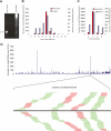
Figure 1.
Figure 1.
A catalog of Chlamydomonas endogenous small RNAs. (A) SYBR-gold was used to visualize small RNAs in total RNA extracted from Chlamydomonas cultures. A 21-nt synthetic RNA oligo was used as a size reference. (B) Size distribution of Chlamydomonas small RNAs. The sets of redundant (red) and unique (blue) small RNAs were used to generate a histogram quantifying the number of sequences obtained for each size class. (C) Sequence composition of the 5′ ends of the small RNAs. (D) Genome-wide density analysis of the small RNAs on an artificial Chlamydomonas genome assembled by linked scaffolds. The number of small RNAs with perfect matches in either a direct or complementary strand within each 50-kb sliding window was plotted. A small RNA production hot spot in Scaffold_58 is shown. The black bar represents the 310-nt hot spot region. Short thin lines above the black bar represent small RNAs derived from the sense strand, and lines below the bar represent small RNAs from the antisense strand. Red lines represent small RNAs that are mapped uniquely to this hot spot, while green lines represent small RNAs that are mapped to multiple genomic sites including the hot spot.
Figure 2.
Figure 2.
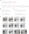
Representative miRNAs in Chlamydomonas. (A–C) Predicted fold-back structures of selected miRNA precursors. Sequences corresponding to the mature miRNAs are shown in red, and sequences corresponding to the miRNA*s are shown in blue. Protruding stem–loops in the 3′ arm of the hairpins are indicated by slashes (\). (D) Confirmation of miRNA expression by Northern blot using end-labeled oligonucleotide probes as indicated. A synthetic 32P-labeled 21-nt RNA oligo was used as a size marker.
Figure 3.
Figure 3.
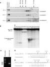
Chlamydomonas miRNAs direct the cleavage of their target mRNAs. (A) Chlamydomonas miRNAs reside in complexes. Extracts from Chlamydomonas cultures were fractionated by size exclusion chromatography. RNAs were extracted from each fraction and used for Northern blotting to detect miRNAs as indicated. The fractions representing 14.4-, 232-, 440-, and 660-kDa size markers, chromatographed separately, are shown. A synthetic 21-nt RNA oligo was used as a size marker. (B) The target of Candidate82 is cleaved in vitro. Each fraction from the size exclusion chromatography was assayed for cleavage activity by incubation with 32P-labeled target transcripts (C_540109, a target of Candidate82). (C) Detection of the 3′ end Candidate82 cleavage product by 5′ RACE assay. A PCR product of the expected size is observed. (D) Identification of the Candidate82 cleavage site. The miRNA target site is aligned with Candidate82. (E) Identification of the Candidate9 cleavage site. The miRNA target site is aligned with Candidate9. Arrows indicate the ends of cleaved mRNA products as determined by 5′ RACE. Above the arrows is shown the frequency of cloned sequences corresponding to each inferred cleavage site. Only cloned sequences that matched the target gene and had a 5′ end within a 50-base-pair (bp) window centered on the miRNA complementary site were counted.
Figure 4.
Figure 4.
Expression patterns of miRNAs in different cell types. The expression of miRNAs in vegetative cells and gametes (CC-621 mt−) was examined by Northern blotting using end-labeled oligonucleotide probes as indicated. Enriched small RNAs prepared from the same number of cells were loaded in each lane. The miRNA signals were quantified and the relative levels in gametes were calculated by comparison with those in vegetative cells (arbitrarily set to 1.0). A synthetic 32P-labeled 21-nt RNA oligo was used as a size marker.
Figure 5.
Figure 5.
Phased siRNA loci in Chlamydomonas. Two representative loci produce siRNAs that are phased to each other. The loci are represented by the long bars at the center of each diagram, and the genomic positions of the loci are shown. Short thin lines above the long bars represent small RNAs derived from the antisense strands, and lines below the bars represent small RNAs from the sense strands. The copy numbers are shown for the small RNAs cloned >50 times.

Comment in

Similar articles

Cited by

References

    1. Allen E., Xie Z., Gustafson A.M., Sung G.H., Spatafora J.W., Carrington J.C., Xie Z., Gustafson A.M., Sung G.H., Spatafora J.W., Carrington J.C., Gustafson A.M., Sung G.H., Spatafora J.W., Carrington J.C., Sung G.H., Spatafora J.W., Carrington J.C., Spatafora J.W., Carrington J.C., Carrington J.C. Evolution of microRNA genes by inverted duplication of target gene sequences in Arabidopsis thaliana. Nat. Genet. 2004;36:1282–1290. - PubMed
    1. Allen E., Xie Z., Gustafson A.M., Carrington J.C., Xie Z., Gustafson A.M., Carrington J.C., Gustafson A.M., Carrington J.C., Carrington J.C. microRNA-directed phasing during trans-acting siRNA biogenesis in plants. Cell. 2005;121:207–221. - PubMed
    1. Aravin A.A., Lagos-Quintana M., Yalcin A., Zavolan M., Marks D., Snyder B., Gaasterland T., Meyer J., Tuschl T., Lagos-Quintana M., Yalcin A., Zavolan M., Marks D., Snyder B., Gaasterland T., Meyer J., Tuschl T., Yalcin A., Zavolan M., Marks D., Snyder B., Gaasterland T., Meyer J., Tuschl T., Zavolan M., Marks D., Snyder B., Gaasterland T., Meyer J., Tuschl T., Marks D., Snyder B., Gaasterland T., Meyer J., Tuschl T., Snyder B., Gaasterland T., Meyer J., Tuschl T., Gaasterland T., Meyer J., Tuschl T., Meyer J., Tuschl T., Tuschl T. The small RNA profile during Drosophila melanogasterdevelopment. Dev. Cell. 2003;5:337–350. - PubMed
    1. Aravin A., Gaidatzis D., Pfeffer S., Lagos-Quintana M., Landgraf P., Iovino N., Morris P., Brownstein M.J., Kuramochi-Miyagawa S., Nakano T., Gaidatzis D., Pfeffer S., Lagos-Quintana M., Landgraf P., Iovino N., Morris P., Brownstein M.J., Kuramochi-Miyagawa S., Nakano T., Pfeffer S., Lagos-Quintana M., Landgraf P., Iovino N., Morris P., Brownstein M.J., Kuramochi-Miyagawa S., Nakano T., Lagos-Quintana M., Landgraf P., Iovino N., Morris P., Brownstein M.J., Kuramochi-Miyagawa S., Nakano T., Landgraf P., Iovino N., Morris P., Brownstein M.J., Kuramochi-Miyagawa S., Nakano T., Iovino N., Morris P., Brownstein M.J., Kuramochi-Miyagawa S., Nakano T., Morris P., Brownstein M.J., Kuramochi-Miyagawa S., Nakano T., Brownstein M.J., Kuramochi-Miyagawa S., Nakano T., Kuramochi-Miyagawa S., Nakano T., Nakano T., et al. A novel class of small RNAs bind to MILI protein in mouse testes. Nature. 2006;442:203–207. - PubMed
    1. Axtell M.J., Bartel D.P., Bartel D.P. Antiquity of microRNAs and their targets in land plants. Plant Cell. 2005;17:1658–1673. - PMC - PubMed

Publication types