. 2007 Jul;75(7):3271-81.
doi: 10.1128/IAI.01810-06. Epub 2007 Apr 30.

Development of fatal colitis in FVB mice infected with Citrobacter rodentium

Affiliations

Development of fatal colitis in FVB mice infected with Citrobacter rodentium

Diana Borenshtein et al. Infect Immun. 2007 Jul.

Abstract

Citrobacter rodentium is the causative agent of transmissible murine colonic hyperplasia. The disease is characterized by severe but temporary epithelial hyperplasia with limited inflammation in the descending colon of adult mice on a variety of genetic backgrounds. The natural history of infection with this murine pathogen has been characterized in outbred Swiss Webster (SW) mice but not in the cognate inbred FVB strain. In contrast to subclinical infection in SW mice, 12-week-old FVB mice developed overt disease with significant weight loss and mortality beginning by 9 days postinoculation (dpi). By 21 dpi, more than 75% of infected FVB mice died or had to be euthanized, whereas no mortality developed in SW mice. Mortality in FVB mice was fully prevented by fluid therapy. Fecal shedding of bacteria was similar in both groups through 9 dpi; however, a slight but significant delay in bacterial clearance was observed in FVB mice by 12 to 18 dpi. SW mice developed hyperplasia with minimal inflammation in the descending colon. FVB mice developed epithelial cell hyperproliferation, severe inflammation with erosions and ulcers, and epithelial atypia by 6 dpi in the descending colon. In the majority of surviving FVB mice, colonic lesions, including epithelial atypia, were reversible, although a small percentage (5 to 7%) exhibited chronic colitis through 7 months postinoculation. The existence of susceptible and resistant lines of mice with similar genetic backgrounds will facilitate the identification of host factors responsible for the outcome of infection and may lead to the development of novel strategies for preventing and treating infectious colitis.


Figures

FIG. 1.
FIG. 1.
Experimental inoculation of FVB mice with C. rodentium causes significant mortality beginning by 9 dpi. By 3 wpi only 23% of infected animals remained alive. Survival curves were generated from five independent experiments (n = 41 for control and n = 84 for infected FVB mice). Differences between infected and control animals were significant (P < 0.0001 by the log rank test). There was no mortality in SW mice with (n = 20) or without infection (n = 16).
FIG. 2.
FIG. 2.
C. rodentium infection in FVB mice results in body weight loss and delayed bacterial clearance. (A) Body weight loss was greater in FVB mice than in SW mice beginning by 9 dpi (P < 0.0001 by two-way ANOVA). Shown are means ± SEM of percent body weight change compared with initial body weight for surviving mice at each time point, which may overestimate the 21-dpi body weight change for FVB mice (see text). *, P < 0.05 for SW and P < 0.0001 for FVB mice by the paired t test. (B) Infection kinetics were similar in both groups through 9 dpi, but bacterial clearance was delayed in FVB mice thereafter. Shown are means ± SEM of log 10-transformed CFU/g of feces for surviving animals with feces at each time point, which may underestimate the fecal shedding of C. rodentium in FVB mice (see text). *, P < 0.05 for 12 dpi and P < 0.0005 for 15 and 18 dpi by Student's t test.
FIG. 3.
FIG. 3.
Colon and cecum counts of C. rodentium do not correlate with bacterial attachment in FVB mice. (A) Tissue homogenates were plated at 3, 6, and 12 dpi. Shown are means ± SEM of log 10-transformed CFU C. rodentium/g of tissue. (B) Although high C. rodentium counts were found at all time points in both the cecum and colon, there was little correlation with attachment of bacteria to epithelial cells identified by IHC for C. rodentium (original magnification, ×200; a, c, and e are colon; b, d, and f are cecum). No C. rodentium was present in control colon or cecum of FVB mice (a and b). Bacterial attachment was observed in the distal colon beginning by 6 dpi. Bacteria are on the luminal surface, as well as deeper in the crypts (c). Although the lumen of the cecum contains bacteria, no attachment to epithelium was observed in the cecum at 6 dpi (d). By 12 dpi, most of the positive signal in the colon was associated with exfoliated cells (e), though some surface attachment was identified in more proximal regions of the colon (not shown). On the other hand, there were occasional areas of focal bacterial attachment to epithelial cells in the cecum at 12 dpi (f).
FIG. 4.
FIG. 4.
C. rodentium infection causes epithelial hyperplasia in the colon of FVB mice. By 6 dpi, there was an increase in crypt column heights and labeling index (A) and expansion of the proliferative zone (B) in the distal colon of infected animals. Shown are mean ± SEM values. *, P < 0.0001 by ANOVA with post hoc Student's t test comparisons between the groups (*, 6 dpi versus 0 and 3 dpi; **, 12 dpi versus 0, 3, and 6 dpi). BrdU labeling in control (C) and 12-dpi colon (D) reveals extensive proliferation of enterocytes throughout the entire crypt including the surface epithelium in infected mice. Original magnification, ×100.
FIG. 5.
FIG. 5.
FVB mice infected with C. rodentium developed colonic inflammation by 6 dpi that was more pronounced by 12 dpi (A). This was accompanied by an increase in epithelial dysplasia (B) (P < 0.0001 using Kruskal-Wallis nonparametric test with subsequent Dunn's multiple comparison test). Each symbol represents one animal. Median lines are presented. Groups indicated by different letters (a and b) are significantly different. Correlation analysis of inflammation versus dysplasia revealed a strong association between these two factors (Spearman r = 0.871; P < 0.0001).
FIG. 6.
FIG. 6.
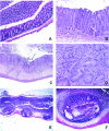
Microscopic lesions in the colon of FVB mice 12 to 15 dpi. (A) Normal colon from an uninoculated FVB mouse. (B) Hyperplastic crypts with increased numbers of mitotic figures and goblet cell depletion. Note significant infiltrate of inflammatory cells in the mucosa and submucosa. (C) Colitis and ulcer (arrow) with marked transmural inflammation and submucosal edema. (D) Epithelial atypia characterized by loss of normal tissue architecture; epithelial cell pleomorphism; and gland malformation with splitting, branching, and infolding. Some mice with more advanced lesions displayed a high nucleus-to-cytoplasm ratio, crypt branching with irregular buds, and occasional micronests within the abutting lamina propria. (E and F) Atypia in gut-associated lymphoid tissue showing herniated dysplastic crypts and dilated and attenuated cysts in submucosa, visualized with hematoxylin and eosin staining. Original magnifications, ×25 (E), ×40 (C), ×100 (A and F), ×200 (B), and ×400 (D).
FIG. 7.
FIG. 7.
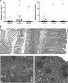
A small percentage of mice develop persistent lesions in the large intestine 16 or 30 wpi with C. rodentium. (A) Lesion scores in the colon and cecum of uninoculated mice and mice at 16 and 30 wpi. Each dot represents one animal. The colonic disease score was slightly, but significantly, greater at 16 wpi (*, P < 0.0001 by Kruskal-Wallis followed by Dunn's multiple comparison test) compared with uninfected controls and mice at 30 wpi. (B) Photomicrographs of chronic lesions. At 16 wpi, inflamed and hyperplastic tissue, mainly in the proximal colon, was segmentally distributed (see adjacent normal mucosa). At 30 wpi, diffuse lesions were observed throughout the colon and cecum. Note the numerous lymphoid aggregates, crypt abscesses, and dilation. Original magnification in panel B, ×200 (a) and ×100 (b and c).
FIG. 8.
FIG. 8.
Quantitative RT-PCR for expression of immunomodulatory cytokine genes. Susceptible FVB and resistant SW mice demonstrated comparable expression of proinflammatory and immunomodulatory genes in response to C. rodentium 9 dpi. Each symbol represents one animal. Horizontal lines indicate mean values.

References

    1. Barthold, S. W. 1979. Autoradiographic cytokinetics of colonic mucosal hyperplasia in mice. Cancer Res. 39:24-29. - PubMed
    1. Barthold, S. W. 1980. The microbiology of transmissible murine colonic hyperplasia. Lab. Anim. Sci. 30:167-173. - PubMed
    1. Barthold, S. W., and A. M. Jonas. 1977. Morphogenesis of early 1,2-dimethylhydrazine-induced lesions and latent period reduction of colon carcinogenesis in mice by a variant of Citrobacter freundii. Cancer Res. 37:4352-4360. - PubMed
    1. Barthold, S. W., and D. Beck. 1980. Modification of early dimethylhydrazine carcinogenesis by colonic mucosal hyperplasia. Cancer Res. 40:4451-4455. - PubMed
    1. Barthold, S. W., G. L. Coleman, P. N. Bhatt, G. W. Osbaldiston, and A. M. Jonas. 1976. The etiology of transmissible murine colonic hyperplasia. Lab. Anim. Sci. 26:889-894. - PubMed

Publication types

MeSH terms

LinkOut - more resources