. 2007 Jul;75(7):3594-603.
doi: 10.1128/IAI.01945-06. Epub 2007 Apr 30.

The Neisseria meningitidis capsule is important for intracellular survival in human cells

Affiliations

The Neisseria meningitidis capsule is important for intracellular survival in human cells

Maria Rita Spinosa et al. Infect Immun. 2007 Jul.

Abstract

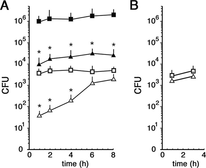
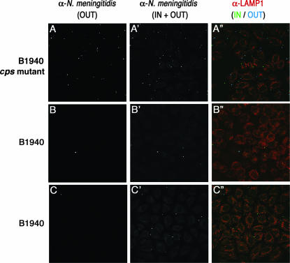
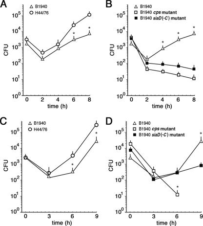
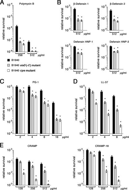
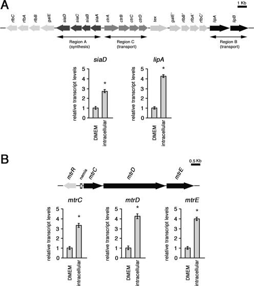
While much data exist in the literature about how Neisseria meningitidis adheres to and invades human cells, its behavior inside the host cell is largely unknown. One of the essential meningococcal attributes for pathogenesis is the polysaccharide capsule, which has been shown to be important for bacterial survival in extracellular fluids. To investigate the role of the meningococcal capsule in intracellular survival, we used B1940, a serogroup B strain, and its isogenic derivatives, which lack either the capsule or both the capsule and the lipooligosaccharide outer core, to infect human phagocytic and nonphagocytic cells and monitor invasion and intracellular growth. Our data indicate that the capsule, which negatively affects bacterial adhesion and, consequently, entry, is, in contrast, fundamental for the intracellular survival of this microorganism. The results of in vitro assays suggest that an increased resistance to cationic antimicrobial peptides (CAMPs), important components of the host innate defense system against microbial infections, is a possible mechanism by which the capsule protects the meningococci in the intracellular environment. Indeed, unencapsulated bacteria were more susceptible than encapsulated bacteria to defensins, cathelicidins, protegrins, and polymyxin B, which has long been used as a model compound to define the mechanism of action of CAMPs. We also demonstrate that both the capsular genes (siaD and lipA) and those encoding an efflux pump involved in resistance to CAMPs (mtrCDE) were up-regulated during the intracellular phase of the infectious cycle.


Figures

FIG. 1.
FIG. 1.
Adherence to and invasion of HeLa cells by N. meningitidis strains. (A) Adherence and invasion assays without centrifugation. HeLa cells were infected with bacteria for the indicated times. The amounts of internalized B1940 (open triangles) and B1940 cps mutant (open squares) bacteria were quantified after saponin treatment. The amounts of adherent B1940 (closed triangles) and B1940 cps mutant (closed squares) bacteria were quantified by omitting the gentamicin step. (B) Invasion assays with centrifugation. The numbers of internalized B1940 (open triangles) and B1940 cps mutant (open squares) bacteria after starting the infection by centrifugation are shown. In A and B, values are means of at least 10 independent experiments made with triplicate samples with standard errors. Asterisks mark statistically significant differences in adherence and invasion values between B1940 and the B1940 cps mutant (P value of <0.05).
FIG. 2.
FIG. 2.
Immunofluorescence analysis of cells infected with the B1940 cps mutant or B1940. HeLa cells were infected with the B1940 cps mutant or B1940 as shown. Images were taken after gentamicin treatment for the B1940 cps mutant (A, A′, and A′′) or after gentamicin treatment (B, B′, and B′′) or 8 h after gentamicin treatment (C, C′, C′′) for B1940. To distinguish between extracellular and intracellular bacteria, the anti-N. meningitidis antibody (α-N. meningitidis) and its secondary antibody were used before (OUT) (A, B, and C) or after (IN + OUT) (A′, B′, and C′) permeabilization of cells with saponin. The secondary antibody used before permeabilization was Cy5 conjugated, while the one used after permeabilization was fluorescein isothiocyanate conjugated. To detect a cellular marker, we used anti-Lamp1 followed by a tetramethyl rhodamine isothiocyanate-conjugated secondary antibody. Merged images of different channels are shown in A′′, B′′, and C′′. Bars, 10 μm.
FIG. 3.
FIG. 3.
Survival/growth of meningococcal strains in HeLa and THP-1 cells. (A and B) HeLa cells (105 cells /well) were infected with different N. meningitidis strains (H44/76 [open circles], B1940 [open triangles], the B1940 cps mutant [open squares], and the B1940 siaD(−C) mutant [closed squares]) at an MOI of 50, treated with gentamicin, and reincubated in DMEM for the indicated times. Bacteria were centrifuged onto cells to start the infection. After saponin lysis, CFU from intracellular bacteria were scored. Values are means of at least 10 independent experiments made in triplicate with standard errors. Asterisks mark statistically significant differences in the numbers of CFU between H44/76 and B1940 (A) and between B1940 and the B1940 cps/B1940 siaD(−C) mutant (P value of <0.05). (C and D) THP-1 cells (105 cells /well) differentiated with 8 nM phorbol myristic acid for 4 days were infected with different N. meningitidis strains (H44/76 [open circles], B1940 [open triangles], the B1940 cps mutant [open squares], and the B1940 siaD(−C) mutant [closed squares]) at an MOI of 5 and treated as described above. Values are means of at least four independent experiments made in triplicate with standard errors. Asterisks mark statistically significant differences in the numbers of CFU between H44/76 and B1940 (C) and between B1940 and the B1940 cps/B1940 siaD(−C) mutant (P value of <0.05).
FIG. 4.
FIG. 4.
Susceptibilities of encapsulated and unencapsulated meningococcal strains to CAMPs. (A to E) Susceptibilities of B1940 (black columns), the B1940 siaD(−C) mutant (gray columns), and the B1940 cps mutant (white columns) to different concentrations of polymyxin B (A), human defensins (β-defensin 1, β-defensin 2, defensin HNP-1, and defensin HNP-2) (B), porcine protegrin PG-1 (C), human cathelicidin LL-37 (D), and murine cathelicidin-related peptides CRAMP and CRAMP-18 (E). Values are means of three independent experiments made in triplicate with standard errors. Asterisks mark statistically significant differences in survival values between B1940 and the B1940 siaD(−C)/B1940 cps mutant (P value of <0.05).
FIG. 5.
FIG. 5.
Semiquantitative analysis of the siaD, lipA, mtrC, mtrD, and mtrE transcripts in intracellular meningococci. (A) Total RNAs were extracted from meningococci (strain B1940) after 8 h of infection of HeLa cells (intracellular) or control bacteria grown for 8 h in culture medium (DMEM). The amounts of siaD- and lipA-specific transcripts were determined by real-time RT-PCR. The scheme of the genomic region encoding capsular elements is reported above the panel. (B) Total RNAs were extracted as described above (A). The amounts of mtrC-, mtrD-, and mtrE-specific transcripts were determined by real-time RT-PCR. The genetic map of the mtrCDE operon and the regulatory gene mtrR is reported above the panel. In A and B, levels of lipA-, siaD-, mtrC-, mtrD-, and mtrE-specific transcripts were normalized to rho mRNA levels. Transcript levels in control bacteria are arbitrarily assumed to equal 1. Each real-time RT-PCR experiment was repeated five times (with triplicate samples) using distinct cDNA preparations, and means and standard deviations (bars) were determined. Asterisks mark statistically significant differences in transcript levels between intracellular and control bacteria (P value of <0.05).
FIG. 6.
FIG. 6.
Semiquantitative analysis of the lipA, mtrC, mtrD, and mtrE transcripts in meningococci exposed to PG-1 or LL-37. (A and B) Total RNAs were extracted from meningococci grown to the late logarithmic phase and then exposed to lethal concentrations (16 μg ml−1) of PG-1 (A) or LL-37 (B) for 0, 5, and 15 min. The amounts of specific transcripts were determined by real-time RT-PCR. (C and D) Total RNAs were extracted from meningococci grown to late logarithmic phase in the presence or in the absence of sublethal concentrations (4 or 8 μg ml−1) of PG-1 (C) or LL-37 (D). The amounts of the specific transcripts were determined as described above. In A to D, levels of lipA-, mtrC-, mtrD-, and mtrE-specific transcripts were normalized to rho mRNA levels. Transcript levels in control bacteria are arbitrarily assumed to equal 1. Each real-time RT-PCR experiment was repeated three times (with triplicate samples) using distinct cDNA preparations, and means and standard deviations (bars) were determined. Asterisks mark statistically significant differences in transcript levels with respect to control samples (P value of <0.05).

Similar articles

Cited by

References

    1. Billker, O., A. Popp, V. Brinkmann, G. Wenig, J. Schneider, E. Caron, and T. Meyer. 2002. Distinct mechanisms of internalization of Neisseria gonorrhoeae by members of the CEACAM receptor family involving Rac1- and Cdc42-dependent and -independent pathways. EMBO J. 21:560-571. - PMC - PubMed
    1. Bucci, C., A. Lavitola, P. Salvatore, L. Del Giudice, D. Massardo, C. Bruni, and P. Alifano. 1999. Hypermutation in pathogenic bacteria: frequent phase variation in meningococci is a phenotypic trait of a specialized mutator biotype. Mol. Cell 3:435-445. - PubMed
    1. Campos, M., M. Vargas, V. Regueiro, C. Llompart, S. Alberti, and J. Bengoechea. 2004. Capsule polysaccharide mediates bacterial resistance to antimicrobial peptides. Infect. Immun. 72:7107-7114. - PMC - PubMed
    1. Deghmane, A., D. Giorgini, M. Larribe, J. Alonso, and M. Taha. 2002. Down-regulation of pili and capsule of Neisseria meningitidis upon contact with epithelial cells is mediated by CrgA regulatory protein. Mol. Microbiol. 43:1555-1564. - PubMed
    1. Dietrich, G., S. Kurz, C. Hubner, C. Aepinus, S. Theiss, M. Guckenberger, U. Panzner, J. Weber, and M. Frosch. 2003. Transcriptome analysis of Neisseria meningitidis during infection. J. Bacteriol. 185:155-164. - PMC - PubMed

Publication types

MeSH terms

Substances

LinkOut - more resources