. 2007 Jul;27(13):4905-16.
doi: 10.1128/MCB.02396-06. Epub 2007 Apr 30.

RINT-1 serves as a tumor suppressor and maintains Golgi dynamics and centrosome integrity for cell survival

Affiliations

RINT-1 serves as a tumor suppressor and maintains Golgi dynamics and centrosome integrity for cell survival

Xiaoqin Lin et al. Mol Cell Biol. 2007 Jul.

Abstract

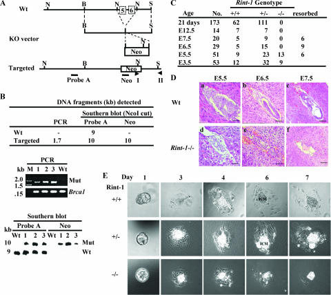
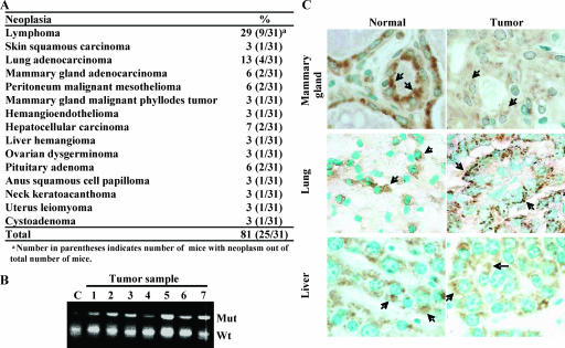
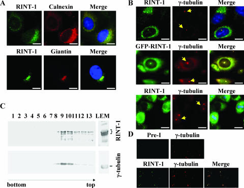
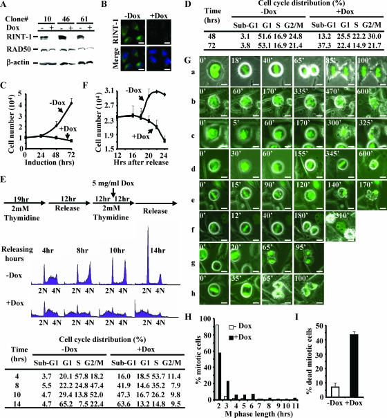
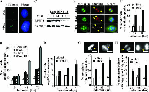
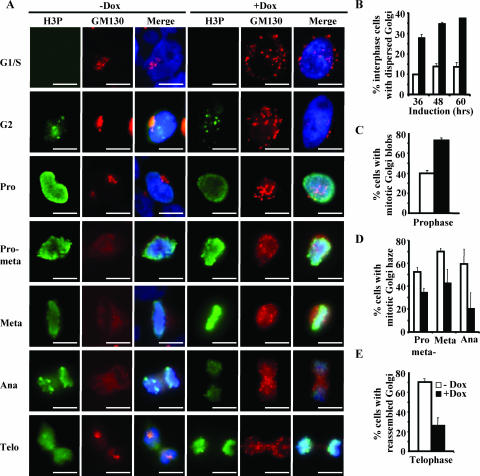
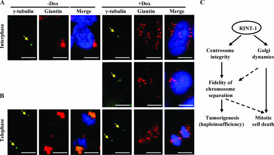
Faithful mitotic partitioning of the Golgi apparatus and the centrosome is critical for proper cell division. Although these two cytoplasmic organelles are probably coordinated during cell division, supporting evidence of this coordination is still largely lacking. Here, we show that the RAD50-interacting protein, RINT-1, is localized at the Golgi apparatus and the centrosome in addition to the endoplasmic reticulum. To examine the biological roles of RINT-1, we found that the homozygous deletion of Rint-1 caused early embryonic lethality at embryonic day 5 (E5) to E6 and the failure of blastocyst outgrowth ex vivo. About 81% of the Rint-1 heterozygotes succumbed to multiple tumor formation with haploinsufficiency during their average life span of 24 months. To pinpoint the cellular function of RINT-1, we found that RINT-1 depletion by RNA interference led to the loss of the pericentriolar positioning and dispersal of the Golgi apparatus and concurrent centrosome amplification during the interphase. Upon mitotic entry, RINT-1-deficient cells exhibited multiple abnormalities, including aberrant Golgi dynamics during early mitosis and defective reassembly at telophase, increased formation of multiple spindle poles, and frequent chromosome missegregation. Mitotic cells often underwent cell death in part due to the overwhelming cellular defects. Taken together, these findings suggest that RINT-1 serves as a novel tumor suppressor essential for maintaining the dynamic integrity of the Golgi apparatus and the centrosome, a prerequisite to their proper coordination during cell division.


Figures

FIG. 1.
FIG. 1.
Targeted disruption of the mouse Rint-1 allele leads to embryonic lethality. (A) Gene-targeting strategy. The region spanning exons 5 and 6 of the Rint-1 gene was replaced with the Neo gene after targeted disruption. Wt, wild type; KO, knockout; B, BamHI; N, NcoI; S, ScaI. Probe A and Neo were probes used for Southern blot analysis. I and II were primers for PCR. (B) Identification of the recombinant ES clones by digesting the genomic DNA with NcoI and probing with probe A or Neo. (Upper panel) Sizes of DNA fragments detected by PCR and Southern blot analyses; middle panel, PCR analysis to confirm the positive clones (the Brca1 gene served as a control); (lower panel) Southern blot analysis to indicate the correct recombinant ES cell clones. M, marker; Mut, mutant; −, none. (C) Genotypic distribution of embryos and mice from Rint-1 heterozygous intercrosses. (D) Histological sections of wild-type and Rint-1/ embryos grown in the uterus. The embryos in the uterus were fixed, sectioned, and stained with hematoxylin and eosin. Rint-1 null embryos manifested abnormal development at E5.5 (d). The resorbed embryos (e and f) were too small to be genotyped and were presumed to be Rint-1/. Bars: panels a and d, 50 μm; panels b, e, and f, 100 μm; and panel c, 200 μm. (E) Blastocysts at E3.5 isolated from Rint-1+/ intercrosses and cultured for 7 days. Wild-type (Rint-1+/+), Rint-1+/, and Rint-1/ blastocysts appeared to be morphologically comparable at day 1. However, while the ICM cells of Rint-1+/+ and Rint-1+/ embryos continued to expand through the 7-day culture period, Rint-1/ ICM cells stopped expanding by day 4 and died later, along with the trophoblastic giant cells.
FIG. 2.
FIG. 2.
Rint-1 heterozygous mice develop multiple tumors. (A) Tumor incidence in Rint-1+/ mice. (B) PCR analysis of microdissected tumor samples showed no loss of heterozygosity in tumors developed in Rint-1+/ mice. C, wild-type mouse embryonic fibroblasts; Mut, mutant; Wt, wild type. (C) Immunostaining with anti-Rint-1 antibody detected the expression of Rint-1 protein (arrows) in mammary, lung, and liver tumors. Specimens were counterstained with methylene green (magnification, ×400).
FIG. 3.
FIG. 3.
RINT-1 is localized to the ER, Golgi apparatus, and centrosome. (A) RINT-1 localized at the ER and Golgi apparatus in U2OS cells. Cells were immunostained with antibodies against RINT-1, calnexin (an ER marker), or Giantin (a Golgi apparatus marker). Bars, 10 μm. (B) RINT-1 localized at the centrosome. (Upper and lower panels) HeLa cells were costained with antibodies against RINT-1 and γ-tubulin, a centrosome marker; (middle panel) GFP-RINT-1 expressed in U2OS cells colocalized with γ-tubulin staining. The arrows indicate the γ-tubulin staining. Bars, 10 μm. (C) Endogenous RINT-1 and γ-tubulin were cofractionated by sucrose density gradient centrifugation. Fractions were immunoblotted with antibodies against either RINT-1 or γ-tubulin. RINT-1 proteins were associated with γ-tubulin through fractions 8 to 13, with a peak in fractions 9 and 10. (D) RINT-1 and γ-tubulin colocalized at the isolated centrosomes. Centrosomes in fractions 9 and 10 were spun onto coverslips and immunostained with preimmune and anti-γ-tubulin antibodies or anti-RINT-1 and anti-γ-tubulin antibodies.
FIG. 4.
FIG. 4.
Depletion of RINT-1 leads to a prolonged M phase and severe mitotic cell death. (A) Characterization of three HeLa cell clones with inducible expression of RINT-1 RNAi. Cells were treated with (+) or without (−) 5 μg of doxycycline/ml for 48 h. The cell lysates were analyzed by Western blotting and probed with antibodies against RINT-1, RAD50, and β-actin. The expression of RINT-1 protein was diminished when RINT-1 RNAi was induced with doxycycline. RAD50 and β-actin served as controls. (B) Confirmation of the depletion of RINT-1 protein in HeLa-RINT-1i cells upon addition of 5 μg of doxycycline/ml for 48 h by immunostaining with anti-RINT-1 antibody. −Dox, without doxycycline; +Dox, with doxycycline. Bars, 10 μm. (C) The depletion of RINT-1 inhibited cell growth of the HeLa-RINT-1i clone. Cells were induced with or without 5 μg of doxycycline/ml, and the viable cells were counted by the trypan blue exclusion assay at the indicated times. Each data point represents the average of results for duplicate samples. The data are representative of results from three independent experiments. Error bars, standard deviations. (D) The depletion of RINT-1 caused cell death in a randomly growing population. HeLa-RINT-1i cells were treated with or without 5 μg of doxycycline/ml for the indicated times, and the cell cycle distribution was analyzed by flow cytometry. A significant increase in sub-G1 cells among RINT-1-depleted cells was observed. The data are representative of results from three independent experiments. (E) RINT-1-depleted cells underwent cell death at the G2/M phase. (Top) Diagram showing the experimental scheme for cell cycle synchronization by double thymidine blocking at the G1/S boundary. (Middle) Profiles from fluorescence-activated cell sorter analysis. Cells were collected and analyzed by flow cytometry. 2N, diploid cells; 4N, tetraploid cells. (Bottom) The percentages of cells at each cell cycle phase are indicated. An increase in the sub-G1 population and a decrease in the G2/M population among RINT-1-depleted cells were simultaneously observed. The data are representative of results from three independent experiments. (F) Proliferation curve of one cell cycle division. HeLa-RINT-1i cells were synchronized at mitotic phase by treatment with 0.1 μg of nocodazole/ml for 8 h. The mitotic cells were collected and replated for 4 h, followed by treatment with or without 5 μg of doxycycline/ml. The viable cells were counted by the trypan blue exclusion assay at the indicated times. Each data point represents the average of results for duplicate samples. The data are representative of results from two independent experiments. Error bars, standard deviations. (G) Living HeLa-RINT-1i cells harboring histone H2B-GFP were monitored under a fluorescence microscope at the indicated times after induction with or without 5 μg of doxycycline/ml for 12 h. Chromosome behavior during M phase was recorded. The mitotic progression of control cells is shown in panel a. The mitotic progression of RINT-1-depleted cells is shown in panels b to h. The numbers indicate the minutes passed during the recording. Bars, 10 μm. (H) The mitotic phase of HeLa-RINT-1i cells expressing histone H2B-GFP and treated with doxycycline was prolonged compared to that of control cells. The M phase length was determined from the intervals between chromosome condensation and the completion of cytokinesis. (I) The percentage of HeLa-RINT-1i/H2B-GFP cells treated with doxycycline that died in the mitotic phase was increased compared to that of control cells. Error bars, standard deviations.
FIG. 5.
FIG. 5.
Depletion of RINT-1 results in centrosome amplification and consequential chromosome instability. (A) Amplified centrosomes in HeLa-RINT-1i cells induced with 5 μg of doxycycline/ml were detected by immunostaining using anti-γ-tubulin (green) and DAPI for the nucleus (blue). +Dox, with doxycycline; −Dox, without doxycycline. Bars, 10 μm. (B) The percentages of cells with centrosome amplification were determined at the indicated times after induction. HeLa-RINT-1i cells were induced with 5 μg of doxycycline/ml and/or 2 mM HU for the indicated time periods. Three hundred cells were counted at each time point. Error bars, standard deviations. (C) The expression of RINT-1 protein in U2OS cells was abolished by infection with rAd-RINT-1i but not with rAd-Luci. Cells were infected with adenoviruses at the indicated multiplicities of infection (MOI) for 72 h, and the cell lysates were analyzed by Western blotting. β-actin served as an internal control. (D) The percentages of U2OS cells infected with rAd-RINT-1i with centrosome amplification were increased compared to those of cells infected with rAd-Luci. Two hundred to 300 cells were counted at each time point. Error bars, standard deviations. (E) Multiple spindles in HeLa-RINT-1i cells were detected upon induction with 5 μg of doxycycline/ml. Cells were coimmunostained with anti-α-tubulin for the mitotic asters and anti-γ-tubulin for the centrosomes (arrows). Bars, 10 μm. (F) The percentages of HeLa-RINT-1i cells with multiple spindles upon doxycycline induction were increased compared to those of control cells. Two hundred cells were counted at each time point. Error bars, standard deviations. (G and H) The percentages of RINT-1-depleted cells with aberrant chromosome segregation, including a misaligned chromosome(s) at metaphase (G) and a lagging or bridging chromosome(s) at telophase (H), were increased compared to those of control cells. The arrows indicate the misaligned, lagging, or bridging chromosomes. Cells at metaphase (n > 200) and anaphase/telophase (n > 200) were examined at each time point. Representative images are shown at the tops of two histograms. All the data are representative of results from two independent experiments. Bars, 10 μm. Error bars, standard deviations.
FIG. 6.
FIG. 6.
Depletion of RINT-1 leads to abnormal Golgi dynamics during cell cycle progression. (A) Micrographs of HeLa-RINT-1i cells coimmunostained with antibodies against GM130, a Golgi apparatus marker, and phosphorylated histone 3 (H3P), which is a marker of different cell cycle stages. Cells were synchronized at the G1/S boundary by double thymidine blocking and induced with (+Dox) or without (−Dox) 5 μg of doxycycline/ml as depicted in Fig. 4E. Pro, prophase; pro-meta, prometaphase; meta, metaphase; ana, anaphase; telo, telophase. Bars, 10 μm. (B to E) The percentages of HeLa-RINT-1i cells with the dispersed Golgi apparatus at interphase (B); mitotic Golgi blob formation at prophase (C); Golgi haze formation at prometaphase, metaphase, and anaphase (D); and the reassembled Golgi structure at telophase (E) after induction with or without 5 μg of doxycycline/ml were determined. Cells at interphase (n > 300); prophase, prometaphase, or metaphase (n > 200); anaphase (n > 50); and telophase (n > 100) were examined. All the data are representative of results from two independent experiments. Error bars, standard deviations.
FIG. 7.
FIG. 7.
Depletion of RINT-1 disrupts the pericentriolar localization of the Golgi apparatus. (A and B) Micrographs of HeLa-RINT-1i cells coimmunostained with antibodies against giantin and γ-tubulin (arrows) upon treating with (+Dox) or without (−Dox) 5 μg of doxycycline/ml. Dissociation of RINT-1 from the pericentriolar region with the dispersed Golgi structure at interphase (A) and with the nonreassembled Golgi structure at telophase (B) was observed in RINT-1-depleted cells (+Dox). Bars, 10 μm. (C) Diagram of proposed RINT-1 functions involved in maintaining centrosome integrity as well as Golgi dynamics. The depletion of RINT-1 leads to mitotic cell death as well as tumorigenesis with haploinsufficiency.

References

    1. Ajiro, K., K. Yoda, K. Utsumi, and Y. Nishikawa. 1996. Alteration of cell cycle-dependent histone phosphorylations by okadaic acid. Induction of mitosis-specific H3 phosphorylation and chromatin condensation in mammalian interphase cells. J. Biol. Chem. 271:13197-13201. - PubMed
    1. Altan-Bonnet, N., R. D. Phair, R. S. Polishchuk, R. Weigert, and J. Lippincott-Schwartz. 2003. A role for Arf1 in mitotic Golgi disassembly, chromosome segregation, and cytokinesis. Proc. Natl. Acad. Sci. USA 100:13314-13319. - PMC - PubMed
    1. Altan-Bonnet, N., R. Sougrat, and J. Lippincott-Schwartz. 2004. Molecular basis for Golgi maintenance and biogenesis. Curr. Opin. Cell Biol. 16:364-372. - PubMed
    1. Arasaki, K., M. Taniguchi, K. Tani, and M. Tagaya. 2006. RINT-1 regulates the localization and entry of ZW10 to the syntaxin 18 complex. Mol. Biol. Cell 17:2780-2788. - PMC - PubMed
    1. Balczon, R., L. Bao, W. E. Zimmer, K. Brown, R. P. Zinkowski, and B. R. Brinkley. 1995. Dissociation of centrosome replication events from cycles of DNA synthesis and mitotic division in hydroxyurea-arrested Chinese hamster ovary cells. J. Cell Biol. 130:105-115. - PMC - PubMed

Publication types

MeSH terms