. 2007 May 7;177(3):539-49.
doi: 10.1083/jcb.200610086. Epub 2007 Apr 30.

Genetic alteration of endothelial heparan sulfate selectively inhibits tumor angiogenesis

Affiliations

Genetic alteration of endothelial heparan sulfate selectively inhibits tumor angiogenesis

Mark M Fuster et al. J Cell Biol. .

Abstract

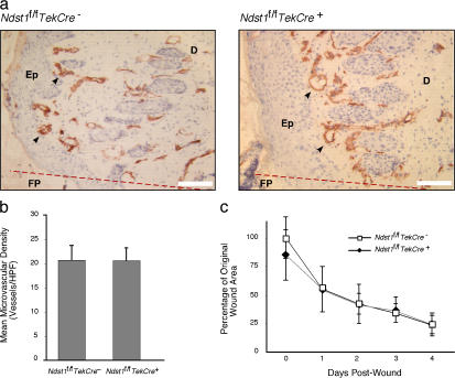
To examine the role of endothelial heparan sulfate during angiogenesis, we generated mice bearing an endothelial-targeted deletion in the biosynthetic enzyme N-acetylglucosamine N-deacetylase/N-sulfotransferase 1 (Ndst1). Physiological angiogenesis during cutaneous wound repair was unaffected, as was growth and reproductive capacity of the mice. In contrast, pathological angiogenesis in experimental tumors was altered, resulting in smaller tumors and reduced microvascular density and branching. To simulate the angiogenic environment of the tumor, endothelial cells were isolated and propagated in vitro with proangiogenic growth factors. Binding of FGF-2 and VEGF(164) to cells and to purified heparan sulfate was dramatically reduced. Mutant endothelial cells also exhibited altered sprouting responses to FGF-2 and VEGF(164), reduced Erk phosphorylation, and an increase in apoptosis in branching assays. Corresponding changes in growth factor binding to tumor endothelium and apoptosis were also observed in vivo. These findings demonstrate a cell-autonomous effect of heparan sulfate on endothelial cell growth in the context of tumor angiogenesis.


Figures

Figure 1.
Figure 1.
Wound healing and angiogenesis is not affected in Ndst1-deficient mice. Mice were subjected to a 3-mm-diameter full-thickness punch lesion on the dorsal skin area. (a) After 4 d, biopsies were taken for immunohistology with CD31 mAb to detect blood vessels in the wound region (arrows). Wounds in both wild-type and mutant mice underwent substantial angiogenesis, as noted by the high density of CD31+ processes detected along the dermis. (b) Density of microvasculature at punch-biopsy wound border, as measured by mean number of CD31+ microvessels per 400× microscopic field surrounding the wound center, is shown for each genotype (mean density calculated from four fields taken about wound center for each mouse; n = 8 mice per group). No significant difference was noted between mutant and wild-type mice (P = 0.96). (c) Contraction of the wound area during healing was measured daily. No significant differences were observed between mutant and wild-type mice (P = 0.288). Anatomic structures in panel a include the wound fibrin plug (FP), located to one side of the original wound margin (represented by the dotted red line), bordered by a proliferating epithelial (Ep) layer and dermis (D). Error bars indicate SD. Bars, 50 μm.
Figure 2.
Figure 2.
Tumor growth is reduced in Ndst1f/fTekCre+ mutant mice. (a) LLC (4 × 10⁵ cells) were injected into the hindquarters of either Ndst1 f/f TekCre + or Ndst1 f/f TekCre mice (n = 5 per group). Tumor volume was estimated by caliper measurements at the indicated times. (b) To control for the genotype of hematopoietic tissue among the various mice, mutant (n = 5) and wild-type (n = 7) mice were transplanted with bone marrow from genotype-matched mice (b, left). A separate group of mutant mice (n = 5) was transplanted with wild-type marrow (b, middle), and a group of wild-type mice was transplanted with mutant bone marrow (b, right). Tumor growth consistently tracked with the genotype of the recipient mouse, indicating that differences in heparan sulfate in the host endothelium were responsible for the altered growth of the tumors. Error bars indicate SD.
Figure 3.
Figure 3.
Visualization of altered tumor microvasculature in Ndst1f/fTekCre+ mice. (a) After 15 d, tumor sections were prepared and stained with CD31 mAb (examples of vessels are indicated by black arrowheads). (b) Vessels were stained with anti-CD31 mAb and visualized by fluorescence microscopy as well. (c) Intravital videomicroscopy of tumor vascular morphology. Representative photomicrographs of tumor spheroid microvasculature 12 d after implantation into skinfold chambers are shown for wild-type (top) and mutant (bottom) spheroids. (d) Representative digitized images of spheroid microvasculature from four independent mice. The first image in each set corresponds to the photomicrographs shown in panel c. (e) Digitized images of four to six regions per chamber were recorded at 100× magnification and scanned and quantified for microvascular branching, grid intersection, linear skeletal length, and caliber. Error bars indicate SD. Bars, 100 μm.
Figure 4.
Figure 4.
Binding of FGF-2 and VEGF164 to Ndst1 mutant microvascular endothelia is reduced. Primary lung microvascular endothelia were isolated from Ndst1 f/f TekCre + or Ndst1 f/f TekCre mice (Materials and methods). (a) Flow cytometry of cells with CD31 mAb showed a high degree of purity. Control incubations contained isotype-matched IgG as primary antibody. (b) Proliferation of mutant versus wild-type primary endothelia in cell culture. Fluorescence in the assay is directly proportional to number of viable cells. Measurements for each time point were performed in triplicate, and the values represent the mean ± SD. (c) The extent of glucosamine N-sulfation, uronyl 2-O-sulfation, or glucosamine 6-O-sulfation was determined by disaccharide analysis of heparan sulfate isolated from mutant and wild-type endothelial cells. (d) Binding of biotinylated FGF-2, VEGF165, and EGF to isolated wild-type (dark gray profiles) and mutant (unfilled black curve) endothelial cells. Controls consisted of cells treated only with streptavidin-FITC (light gray profiles). (e) Equal amounts of growth factor were added to purified heparan [35S]sulfate isolated from mutant and wild-type endothelial cells, and complexes were collected by membrane filtration. Binding of growth factor in the presence of heparin (+Hep) is shown as a control. Error bars indicate SD.
Figure 5.
Figure 5.
Altered sprouting of Ndst1-deficient microvascular endothelial cells on reconstituted extracellular matrix. (a) Wild-type endothelial cells formed extensive networks of interconnected processes when plated on Matrigel with FGF-2 at 10 ng/ml (top). Under the same conditions, sprouting of mutant cells was markedly impaired (bottom). Bar, 200 μm. (b) Process formation by wild-type cells varied with concentration of FGF-2, whereas mutant cells showed essentially no sprouting under these conditions. Treatment with saturating levels of growth factor (100 ng/ml) was sufficient to restore sprouting by mutant cells. (c) Sprouting by wild-type cells to 10 ng/ml FGF-2 was inhibited by the addition of either 3 mU/ml heparinase or 100 μg/ml heparin, whereas the marginal sprouting by mutant cells was unaffected. Data are mean values ± SD for net length of endothelial processes per (100×) microscopic field, normalized to the response of unstimulated wild-type cells. (d) Mutant cells were also insensitive to 10 ng/ml VEGF164. Data are the mean ± SD of three independent experiments normalized to the response of unstimulated wild-type endothelia.
Figure 6.
Figure 6.
Altered growth factor signaling by Ndst1-deficient microvascular endothelia. (a) TUNEL assay was performed on mutant and wild-type endothelial cells undergoing sprouting on Matrigel. In these experiments, cells derived from Ndst1 f/f TekCre mice were infected with Ad-Cre (Ndst1 f/f Ad-Cre) or Ad-GFP virus as a control (Ndst1 f/f Ad-GFP). After infection with Ad-Cre, numerous apoptotic bodies were observed within the cell clusters (bottom, arrows). Ad-GFP–infected cells formed extensive sprouting networks that were relatively free of apoptotic bodies, and the few nonsprouting clusters that were present also were devoid of apoptotic bodies (top right, inset). Bars, 100 μm. (b) Phosphorylation of Erk (p44/p42) in response to the growth factors FGF-2 or VEGF164 was measured by Western blotting. The bands were scanned, the intensity of the phosphorylated protein was normalized to the intensity of the nonphosphorylated protein, and the ratio was scaled to the value obtained at t = 0 (values in parentheses). Total Erk was similar in mutant and wild-type cells. No significant variation in signal intensity >60 min was observed in the absence of growth factor in mutant and wild-type endothelia. (c) Phosphorylation of Akt was measured in wild-type and mutant cells.
Figure 7.
Figure 7.
Altered growth factor binding to tumor microvasculature in Ndst1f/fTekCre+ mice. (a) Binding of biotinylated FGF-2 to histologic sections of LLC tumors. Solid arrowheads in wild-type sections (top) indicate strong binding in perivascular locations (surrounding birefringent red blood cells within vessels) compared with sections from mutant mice (bottom, open arrowheads). (b) Binding of GV39M to VEGF–VEGFR2 complexes and laminin mAb to vessels in tumor sections. In wild-type mice (top), GV39M (red fluorescence) colocalized with vessel-associated laminin (green), as demonstrated in merged images (yellow). Sections from mutant mice (bottom) did not show vascular colocalization of complexes with laminin. (c) Tumor sections from wild-type mice costained with CD31 mAb and GV39M (red) showed that the VEGF–VEGFR complexes were located ablumenal to CD31 staining (green). (d) Tumor sections from wild-type and mutant mice were stained for smooth muscle actin (brown) to assess the degree of vascular investment by pericytes. Mean density of stained processes among tumors in wild-type versus mutant backgrounds was comparable (n = 5 mice per group; P = 0.3). (e) Vascular endothelium-associated apoptosis in tumors was examined by labeling for CD31 (blue) and TUNEL (brown). Vasculature in tumors from mutant mice showed occasional vessel-associated apoptotic bodies (right, arrowheads), whereas any TUNEL reaction in wild-type sections was only associated tumor nuclei (left, arrowhead). (f) Growth of tumors on a VEGFR2 heterozygous background. LLC tumors were grown on the hindquarters of wild type as well as VEGFR2 +/ mice. Tumor volumes at 15 d are shown as scatter plots for wild type (n = 11 mice; solid circles) and VEGFR2 heterozygous mutants (n = 12 mice; open circles), with mean values indicated by horizontal bars on the graph. (g) Tumor microvasculature stained by anti-CD31 mAb (red) in VEGFR2 mutant sections was also altered (right) relative to wild type (left). Bars: (a, d, and g) 50 μm; (b) 15 μm; (c and e) 20 μm.

References

    1. Bai, X., G. Wei, A. Sinha, and J.D. Esko. 1999. Chinese hamster ovary cell mutants defective in glycosaminoglycan assembly and glucuronosyltransferase I. J. Biol. Chem. 274:13017–13024. - PubMed
    1. Bame, K.J., and J.D. Esko. 1989. Undersulfated heparan sulfate in a Chinese hamster ovary cell mutant defective in heparan sulfate N-sulfotransferase. J. Biol. Chem. 264:8059–8065. - PubMed
    1. Bergsland, E.K. 2004. Update on clinical trials targeting vascular endothelial growth factor in cancer. Am. J. Health Syst. Pharm. 61:S12–S20. - PubMed
    1. Bix, G., and R.V. Iozzo. 2005. Matrix revolutions: “tails” of basement-membrane components with angiostatic functions. Trends Cell Biol. 15:52–60. - PubMed
    1. Blackhall, F.H., C.L.R. Merry, E.J. Davies, and G.C. Jayson. 2001. Heparan sulfate proteoglycans and cancer. Br. J. Cancer. 85:1094–1098. - PMC - PubMed

Publication types

MeSH terms

Substances