. 2007 May 7;177(3):451-64.
doi: 10.1083/jcb.200612023. Epub 2007 Apr 30.

Hif-1alpha regulates differentiation of limb bud mesenchyme and joint development

Affiliations

Hif-1alpha regulates differentiation of limb bud mesenchyme and joint development

Sylvain Provot et al. J Cell Biol. .

Abstract

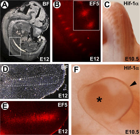
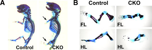
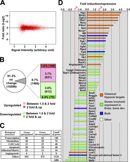
Recent evidence suggests that low oxygen tension (hypoxia) may control fetal development and differentiation. A crucial mediator of the adaptive response of cells to hypoxia is the transcription factor Hif-1alpha. In this study, we provide evidence that mesenchymal condensations that give origin to endochondral bones are hypoxic during fetal development, and we demonstrate that Hif-1alpha is expressed and transcriptionally active in limb bud mesenchyme and in mesenchymal condensations. To investigate the role of Hif-1alpha in mesenchymal condensations and in early chondrogenesis, we conditionally inactivated Hif-1alpha in limb bud mesenchyme using a Prx1 promoter-driven Cre transgenic mouse. Conditional knockout of Hif-1alpha in limb bud mesenchyme does not impair mesenchyme condensation, but alters the formation of the cartilaginous primordia. Late hypertrophic differentiation is also affected as a result of the delay in early chondrogenesis. In addition, mutant mice show a striking impairment of joint development. Our study demonstrates a crucial, and previously unrecognized, role of Hif-1alpha in early chondrogenesis and joint formation.


Figures

Figure 1.
Figure 1.
Mesenchymal condensations are hypoxic, and Hif-1α is expressed in limb bud mesenchyme. The EF5 staining (B and E) and corresponding bright-field (BF) views (A and D) of developing axial skeleton (A and B) and forelimb (D and E) of E12 mouse embryo show the hypoxic tissues. The boxed area (developing axial skeleton) and the area outlined by a dashed line (presumptive digital ray) indicate the regions shown for the EF5 staining. A blow-up of two hypoxic mesenchymal condensations is shown in B. Whole mount immunohistochemistry for Hif-1α protein (C and F), on E10.5 mouse embryo shows that Hif-1α protein is detected in the somites, giving rise to the axial skeleton (C) in limb bud mesenchyme (F, asterisk) and in the apical ectoderm (F, arrowhead).
Figure 2.
Figure 2.
HREs are activated in condensed mesenchyme. Five HREs were placed before a minimal promoter (mp) fragment driving the LacZ reporter gene to generate a hypoxia-inducible (5XHRE-mp/LacZ) transgenic reporter mouse (A). X-gal staining of E8.5 5XHRE-mp/LacZ transgenic embryos cultured ex vivo under normoxic (21% oxygen) or anoxic (0% oxygen) conditions for 24 h indicates a good response of the construct to hypoxia (B; h, heart; ys, yolk sac). Whole mount X-gal staining on 5XHRE-mp/LacZ transgenic embryos of the indicated stage shows the transcriptional activation of HREs in somites (C), developing axial skeleton (D), and limb bud mesenchyme (F), and shows a particularly strong activation in condensed limb mesenchyme (G). The pink circles in F define the margins of the limb bud. Sections of the developing axial skeleton (E) and condensed limb mesenchyme (H) show that the X-gal staining closely resembles that obtained with EF5 (Figure 1).
Figure 3.
Figure 3.
Conditional removal of Hif-1α in limb bud mesenchyme leads to limb deformities. (A) Skeletal preparations of newborn control and CKO mice show the characteristic shortening of CKO limbs (arrows). (A) Skeletal preparations of forelimbs (FL) and hindlimbs (HL) of control and CKO newborn littermates.
Figure 4.
Figure 4.
Hif-1α is dispensable for condensation of limb bud mesenchyme. PNA staining (A and B), hematoxylin and eosin (H&E) staining (C and D), and in situ hybridization with the indicated probes (E–H) on the autopod of E12.5 forelimbs indicate that limb mesenchymal condensations form normally in the absence of Hif-1α.
Figure 5.
Figure 5.
Hif-1α is required for early chondrocyte differentiation. H&E staining (A and B) of E13.5 forelimb autopod, and Alcian blue staining (C and D) of E13.5 distal portion of forelimb autopod, indicate an abnormal histology of the cartilage and an abnormal cartilaginous matrix production, respectively. At this stage, a blow up of cells present in the cartilaginous primordia indicates the presence of differentiated cuboidal chondrocytes in control (E) and undifferentiated mesenchymal cells in CKO autopod (F). In situ hybridization reveals that E13.5 CKO metacarpals (H) express slightly less Col2a1 mRNA at this stage than control (G). In situ hybridization with the indicated markers performed on E14.5 forelimb control and CKO autopods confirms the dramatic delay in chondrocyte differentiation in the absence of Hif-1α (I–P).
Figure 6.
Figure 6.
Joints are hypoxic and express Hif-1α. (A) Immunohistochemistry for Hif-1α on E14.5 forelimb autopod shows a particularly strong expression in the joints of the digits (arrows) and in the articular region of the wrist (arrowheads), in addition to the hypertrophic chondrocytes (asterisk). This expression is significantly reduced in CKO littermates (A′). In situ hybridization with a VEGF probe (B) closely resembles the Hif-1α protein expression pattern. EF5 staining indicates that the forming joints in the digits (C) and the articular region in the wrist (D) are highly hypoxic in E13.5 autopods. H&E staining indicates that the perichondrium surrounding the joints is particularly thick (E). Chondrocytes present at the articular surface remain hypoxic after joint cavitation, as indicated by EF5 staining of metacarpus (F) and elbow (G) of E15.5 forelimbs.
Figure 7.
Figure 7.
Hif-1α is required for joint development. H&E staining of E14.5 (A and B) and newborn (NB; C and D) forelimb autopod shows a delay in joint development in absence of Hif-1α. In situ hybridization with a GDF5 probe on E13 (E and F) and E14.5 (G and H) forelimb autopods indicates the absence of joint specification (F) and the subsequent abnormal joint development (H) in the absence of Hif-1α. The absence of some metacarpals in D results from an artifact of sectioning.
Figure 8.
Figure 8.
CKO of Hif-1α in limb bud mesenchyme alters the formation of wrists. H&E staining of newborn (NB; A and B) and P9 (C and D) wrists and skeletal preparation of P9 wrists (E and F) show that bones present in the CKO wrist are misshapen, dislocated, and partially fused together for some of them. Wrist bones are indicated on each section. 1–3 and 4/5, distal row of carpal bones; c, central carpal bone; r and u, radial and ulnar bones; Ra and Ul, radius and ulna. Slashes between bone designators (i.e., 4/5) indicate a fusion of the indicated bones. 4/5, but never c/3, are always fused in wild-type wrist.
Figure 9.
Figure 9.
Global effect of hypoxia on gene expression in cartilage. (A) A microarray analysis of the genes expressed in metatarsal explants and regulated upon 8 h of hypoxic treatment shows a symmetrical distribution of up- versus down-regulated genes. (B) The relative percentages of genes regulated in function of their fold induction or repression is indicated. Only the genes found to be at least 1.5-fold induced or repressed with a good P-value were considered to be regulated (see Materials and methods for all of the parameters used to filter the genes). (C) Genes regulated upon 8 h of hypoxia are involved in many diverse biological processes. A selection of a few regulated genes and their respective fold induction or repression is given in D. The genes located between −1.5- and +1.5-fold are not considered significantly regulated.
Figure 10.
Figure 10.
Hif-1α regulates early chondrogenesis and joint development. TUNEL assay (A and B) performed on E14.5 forelimb autopods does not reveal any significant cell death in the absence of Hif-1α (B) in this tissue and at this stage. (C–F) In situ hybridization using a VEGFR2 probe (which marks endothelial cells) on E12.5 (C and D) and E14.5 (E and F) forelimbs does not indicate any detectable angiogenic problem in CKO autopod (G). Lack of Hif-1α alters differentiation in the absence of cell death and without impairing angiogenesis. A model for Hif-1α regulation of early chondrocyte differentiation and joint development.

References

    1. Akiyama, H., M.C. Chaboissier, J.F. Martin, A. Schedl, and B. de Crombrugghe. 2002. The transcription factor Sox9 has essential roles in successive steps of the chondrocyte differentiation pathway and is required for expression of Sox5 and Sox6. Genes Dev. 16:2813–2828. - PMC - PubMed
    1. Bi, W., J.M. Deng, Z. Zhang, R.R. Behringer, and B. de Crombrugghe. 1999. Sox9 is required for cartilage formation. Nat. Genet. 22:85–89. - PubMed
    1. Bi, W., W. Huang, D.J. Whitworth, J.M. Deng, Z. Zhang, R.R. Behringer, and B. de Crombrugghe. 2001. Haploinsufficiency of Sox9 results in defective cartilage primordia and premature skeletal mineralization. Proc. Natl. Acad. Sci. USA. 98:6698–6703. - PMC - PubMed
    1. Bishop, T., K. Lau, A. Epstein, S. Kim, M. Jiang, D. O'Rourke, C. Pugh, J. Gleadle, M. Taylor, J. Hodgkin, and P. Ratcliffe. 2004. Genetic analysis of pathways regulated by the von hippel-lindau tumor suppressor in Caenorhabditis elegans. PLoS Biol. 2:e289. - PMC - PubMed
    1. Brunet, L.J., A. McMahon, and R.M. Harland. 1998. Noggin, cartilage morphogenesis, and joint formation in the mammalian skeleton. Science. 280:1455–1457. - PubMed

Publication types

Substances