. 2006:104:453-77.

Integrins in the optic nerve head: potential roles in glaucomatous optic neuropathy (an American Ophthalmological Society thesis)

Affiliations

Integrins in the optic nerve head: potential roles in glaucomatous optic neuropathy (an American Ophthalmological Society thesis)

John C Morrison. Trans Am Ophthalmol Soc. 2006.

Abstract

Purpose: To demonstrate that specific distributions of integrin-based focal mechanoreceptors exist in primate optic nerve heads, suitable for translating stress and strain into the cellular responses of glaucomatous optic neuropathy.

Methods: Normal human (N = 20) and rhesus monkey (N = 14) optic nerve heads and 32 glaucomatous optic nerve heads were processed for immunohistochemistry to determine the structural distribution of integrin subunits alpha1, alpha2, alpha3, alpha4, alpha5, alpha6, alphav, beta1, beta2, beta3, and beta4. Labeling patterns in glaucoma specimens were compared with those of normal eyes.

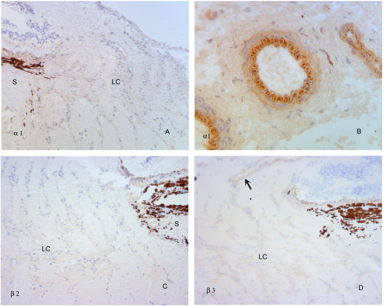
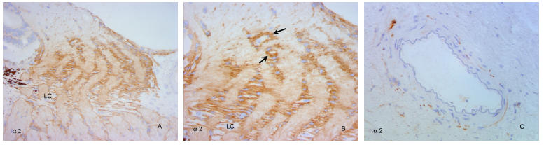
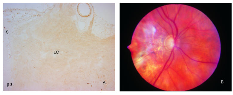
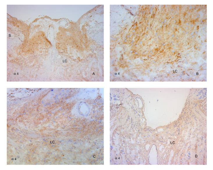
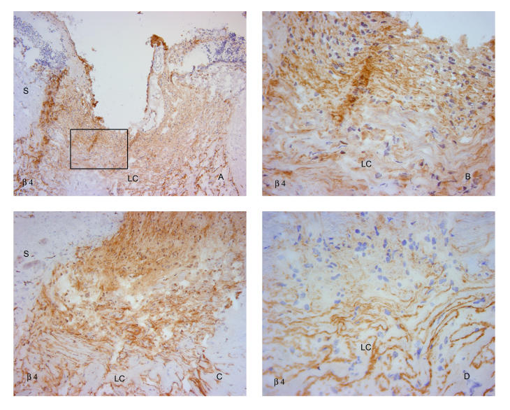
Results: In all specimens, cells within collagenous laminar beams and sclera failed to label with any integrin antibodies. In normal eyes, alpha2, alpha3, alpha6, beta1, and beta4 antibodies localized to astrocytes along the margins of laminar beams and within glial columns. alpha3, alpha5, alpha6, alphav, beta1, and beta4 labeled vascular endothelial cells. In severely damaged glaucoma specimens, cells anterior to the compressed lamina cribrosa displayed persistent label for alpha2, alpha3, beta1, and beta4, whereas label for alpha4 increased and alpha6 was decreased.

Conclusions: Integrins alpha2beta1, alpha3beta1, alpha6beta1, and alpha6beta4 may provide attachment for astrocytes to basement membranes via laminin, providing opportunities to sense changes in stress and strain within and anterior to the lamina cribrosa. Vascular endothelial cell stress may be mediated by integrins alpha3beta1, alpha6beta1, and alpha6beta4, along with alpha5beta1 and alphavbeta1. In advanced damage, reduced alpha6 label and variable label for betabeta 4 anterior to the lamina cribrosa suggests astrocyte migration. Increased label for alpha4 subunits suggests activation of microglia.


Figures

FIGURE 1
FIGURE 1
Ultrastructure of the optic nerve head. A, Astrocyte glial processes (arrows) demonstrate intimate relationships to optic nerve axons (cut in cross section). B, Within laminar beam, fibrillar collagen (cut in cross section and longitudinally) is interposed between capillary and astrocytes lining the beam itself. Note basement membranes (arrowheads) of capillary endothelial cells and astrocytes. As, astrocytes; Cap, capillary. (Magnification ×40,000)
FIGURE 2
FIGURE 2
Distribution of common ECM integrin ligands within the normal primate optic nerve head. Antibodies to fibrillar collagens 1 (A) and 3 (B) demonstrate dense label of the collagenous inner portions of the laminar beams, with little extension into the anterior region of the optic nerve head, except in association with blood vessels (arrow). Note lack of label in association with individual astrocytes in this region. Collagen 4 (C) and laminin (D) antibodies label the margins of the laminar beams, (*) corresponding to basement membranes of the astrocyte foot processes. In contrast to the fibrillar collagens, both of these constituents, and in particular laminin, appear anterior to the collagenous lamina, tapering into the glial columns in association with blood vessels and astrocytes (arrowheads) (D and E). Fibronectin (F), which generally colabels with fibrillar collagens, also labels the collagenous laminar beams as well as peripapillary sclera and major blood vessels, and is present over the glial columns anteriorly. Tenascin (G) demonstrates a very distinct label of the sclera (S) and peripapillary sclera, but not the optic nerve sheath (Sh) or pia (P), in distinction to the fibrillar collagens. Within the lamina cribrosa (*), tenascin labels the laminar beams (H and I), but this does not extend anteriorly (arrowhead), except in association with blood vessels (arrow). Higher-power view (I) illustrates label of laminar beams, without cellular label in between the individual beams. Label with vitronectin antibodies (J) was heavier over the anterior portions of the lamina and glial columns, with relatively less intense staining of the collagenous laminar beams (*). (Magnification: A, ×68; B, ×136; C, ×68; D, ×272; E, ×272; F, ×272; G, ×17; H, ×68; I, ×272; J, ×136)
FIGURE 3
FIGURE 3
α1, β2, and β3 integrins within the normal primate optic nerve head. Antibodies to α1 integrin demonstrate none to minimal specific label (A), with the exception of larger blood vessels of the peripapillary sclera (B). β2 antibodies (C) failed to demonstrate label to any identifiable structure of the optic nerve head. β3 antibodies (D) also failed to produce specific label, with the exception of large retinal vessel walls (arrow). S, sclera; LC, lamina cribrosa. (Magnification: A, ×136; B, ×272, C, ×136, D, ×136)
FIGURE 4
FIGURE 4
α4, α5 and αv integrins within the normal primate optic nerve head. Antibodies to α4 integrin (A) demonstrate only scattered label (arrows) within the laminar and prelaminar optic nerve head, without labeling an arteriole in the peripapillary sclera (arrowhead). Higher-power views of the lamina (B, C) illustrate discontinuous label of small vessels (arrows). α5 (D) and αv (E) antibodies produce strong label of the central retinal vessels and capillaries of the lamina cribrosa and anterior optic nerve head. High-power view (F) documents a continuous label pattern in contrast to the discontinuous pattern shown by α4 integrin. Similar findings were observed for αv (not shown). Label of laminar beam margins and glial columns was absent or minimal with all of these antibodies. (Magnification: A, ×136, B, ×272, C, ×680; D, ×136; E, ×272; F, ×680)
FIGURE 5
FIGURE 5
α2 integrins within the normal primate optic nerve head. α2 integrin antibodies (A) demonstrate label of astrocytes surrounding the collagenous laminar beams, with increased intensity over the glial columns just anterior to the laminar beams. Higher-power view (B) shows label around, but not within, laminar beams at lower part of micrograph, with label of astrocyte columns extending anteriorly, into the lamina choroidalis. Collections of astrocytes outline small blood vessels (arrows), which otherwise are not specifically labeled in the nerve fiber layer region of the optic nerve head. (C) Peripapillary arteriole not labeled with antibodies to α2. LC, lamina cribrosa. (Magnification: A, ×136; B, ×272; C, ×272)
FIGURE 6
FIGURE 6
α3, α6, β1, and β4 integrin distribution within the normal primate optic nerve head is similar to that of α2, with the exception of the vasculature. Low-power micrographs (A and B) illustrate overall label of laminar beam margins, with heavier label over the glial columns, tapering anteriorly into the nerve fiber layer. Note label of vasculature in anterior optic nerve head (arrows). Higher-power views document label of laminar beam margins (C) and transition from laminar beams to glial columns (D, E, F). All antibodies labeled inner margins of blood vessels and capillaries within the lamina cribrosa (G) and those anterior to the lamina, extending into the nerve fiber layer (H). S, sclera; LC, lamina cribrosa. (Magnification: A, ×136; B, ×136; C, ×272; D, ×272; E, ×136; F, ×272; G, ×680; H, 272)
FIGURE 7
FIGURE 7
ECM integrin ligands in glaucomatous optic nerve damage. Collagen 1 label in an eye with severe optic nerve damage from primary open-angle glaucoma (A and B). Note condensed lamina cribrosa (LC), with loss of normal layered architecture and unlabeled cellular layer anterior to the lamina. Tenascin antibody label (C and D) is primarily restricted to the compressed lamina cribrosa in a similarly damaged eye, with relatively little cellular label within, and anterior to the laminar beams. Vitronectin label appears most intense (arrows) over the anterior laminar beams in an eye with primary open-angle glaucoma (E) and neovascular glaucoma (F). Arrowhead in F indicates less intense label of posterior lamina cribrosa. S, sclera. (Magnification: A, ×68; B, ×272; C, ×68; D, ×272; E, ×272; F, ×272)
FIGURE 8
FIGURE 8
Label of eye with moderate glaucomatous optic nerve damage using antibodies to β1 integrin. Note general disorganization of normal lamellar structure and poor delineation of lamina cribrosa (LC). (Magnification ×68)
FIGURE 9
FIGURE 9
α1, β2, and β3 integrin labeling of cells in the optic nerve head is not altered in glaucoma. As illustrated for β3 (A), antibodies to these integrins do not specifically label lamina cribrosa (LC) region of the optic nerve head from an eye with extensive optic nerve damage. Note labeling of central retinal vessel at top of photograph, as also observed in normal eyes. A clinical photograph of this eye is shown in (B). S, sclera; LC, lamina cribrosa. (Magnification: A, ×68)
FIGURE 10
FIGURE 10
Vascular labeling with αV and α5 integrin antibodies is not altered in glaucomatous optic nerve heads. Antibodies to αv (A) and α5 (B) produced expected label of blood vessels only (arrows) in optic nerve head from specimen shown in Figure 9. Similar pattern with antibodies to α5 in another specimen with advanced glaucomatous injury is seen in C. Label of cells anterior to the compressed lamina cribrosa is nearly absent. LC, lamina cribrosa. (Magnification: A, ×68; B, ×272; C, ×272)
FIGURE 11
FIGURE 11
Glaucomatous damage intensifies α4 labeling anterior to the lamina cribrosa. Antibodies to α4 reveal intensified label of cells anterior to the lamina cribrosa in an eye with severe nerve damage from neovascular glaucoma (A and B) and in a specimen with primary open-angle glaucoma (C), also shown in Figure 9C. However, in a third eye with advanced cupping from primary open-angle glaucoma (D), label intensity was not dramatically increased. Label intensity of cells within the lamina cribrosa was relatively minimal and not noticeably altered from that in normal tissues. S, sclera; LC, lamina cribrosa. (Magnification: A, ×68; B, ×272; C, ×136; D, ×68)
FIGURE 12
FIGURE 12
Glaucomatous damage does not affect prelaminar labeling with α3 and β1 integrin antibodies but decreases label intensity for α6 integrin. Comparative label with antibodies to α3 (A and B), β1 (C and D), and α6 (E and F) in optic nerve head from two glaucomatous eyes, one with extensive damage from primary open-angle glaucoma (A, C, E) and one with neovascular glaucoma (B, D, F). α3 and β1 showed strong label of cells anterior to and between the compressed, posteriorly bowed lamina cribrosa in both specimens. In contrast, α6 label of prelaminar cells was surprisingly weak, whereas that between the laminar beams was similar to that in normal eyes. S, sclera; LC, lamina cribrosa. (Magnification: A, ×68; B, ×136; C, ×68; D, ×136; E, ×68; F, ×136)
FIGURE 13
FIGURE 13
Prelaminar labeling with α2 and α3 antibodies in glaucomatous eyes is unaffected, in contrast to diminished intensity of α6. Two additional eyes with advanced damage from primary open-angle glaucoma demonstrate heavy cellular label with antibodies to α2 (A and B) and α3 integrin (C and D). α6 antibody label (E) was again reduced, although not completely absent in one of these specimens. LC, lamina cribrosa. (Magnification: A, ×136; B, ×272; C, ×68; D, ×272; E, ×136)
FIGURE 14
FIGURE 14
β4 integrin in glaucomatous eyes. Label with antibodies to β4 demonstrated variable patterns, with heavy anterior cellular labeling in an eye with advanced primary open-angle glaucoma (A and B) and in another with neovascular glaucoma (C). Label was markedly diminished in another with primary open-angle glaucoma damage (D). S, sclera; LC, lamina cribrosa. (Magnification: A, ×68; B, ×272; C, ×136; D, ×272)
FIGURE 15
FIGURE 15
Aggrecan label in normal and glaucomatous eyes. Antibodies to the large chondroitin sulfate proteoglycan aggrecan selectively labels tissues subjected to high stress (A and B), including the sclera and blood vessel walls, but not tissues that experience lower stress, such as the optic nerve sheath (Sh) and the pia mater. Lamina cribrosa demonstrates variable results, with light label (C) of laminar beams in some specimens (same eye as A), and more intense labeling in others (D), with reduced intensity over the more posterior beams (arrows). Eyes with moderate optic nerve damage from primary open-angle glaucoma typically demonstrate heavier label of lamina (E and F), with abnormal deposits (*) between laminar beams. (Magnification: A, ×17; B, ×17; C, ×272; D, ×272; E, ×17; F, ×272)
FIGURE 16
FIGURE 16
Nonhuman primate optic nerve head labeled with antibodies to GFAP. Note increased density of label (A) just anterior to the lamina cribrosa (LC). On higher power (B) this appears to be related to astrocytes of the glial columns and their transverse processes. (Magnification: A, ×17; B, ×136)

References

    1. Friedman DS, Wolfs RC, O’Colmain BJ, et al. Prevalence of open-angle glaucoma among adults in the United States. Arch Ophthalmol. 2004;122:532–538. - PMC - PubMed
    1. Quigley HA. New paradigms in the mechanisms and management of glaucoma. Eye. 2005;19:1241–1248. - PubMed
    1. Tielsch JM, Sommer A, Katz J, et al. Racial variations in the prevalence of primary open-angle glaucoma. The Baltimore Eye Survey. JAMA. 1991;266:369–374. - PubMed
    1. Mitchell P, Smith W, Attebo K, et al. Prevalence of open-angle glaucoma in Australia. The Blue Mountains Eye Study. Ophthalmology. 1996;103:1661–1669. - PubMed
    1. Klein BE, Klein R, Sponsel WE, et al. Prevalence of glaucoma. The Beaver Dam Eye Study. Ophthalmology. 1992;99:1499–1504. - PubMed

Publication types