

A two-dimensional biomechanical model of vocal fold posturing
- PMID: 17471739
- PMCID: PMC6371396
- DOI: 10.1121/1.2697573
A two-dimensional biomechanical model of vocal fold posturing
Abstract
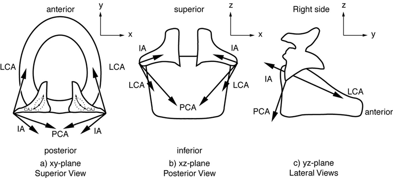
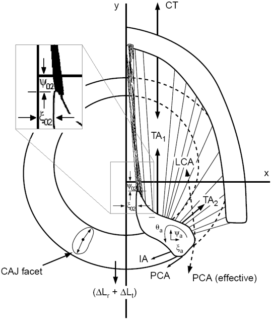
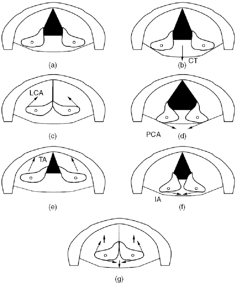
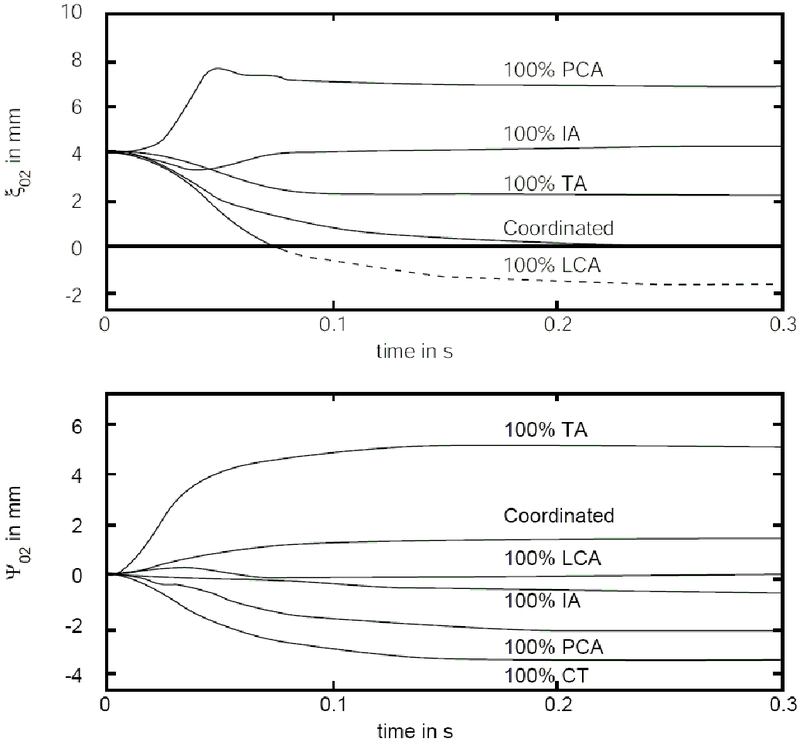
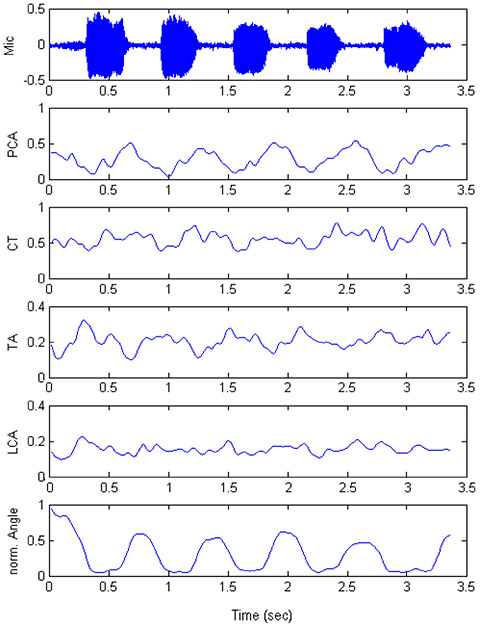
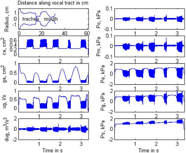
The forces and torques governing effective two-dimensional (2D) translation and rotation of the laryngeal cartilages (cricoid, thyroid, and arytenoids) are quantified on the basis of more complex three-dimensional movement. The motions between these cartilages define the elongation and adduction (collectively referred to as posturing) of the vocal folds. Activations of the five intrinsic laryngeal muscles, the cricothyroid, thyroarytenoid, lateral cricoarytenoid, posterior cricoarytenoid, and interarytenoid are programmed as inputs, in isolation and in combination, to produce the dynamics of 2D posturing. Parameters for the muscles are maximum active stress, passive stress, activation time, contraction time, and maximum shortening velocity. The model accepts measured electromyographic signals as inputs. A repeated adductory-abductory gesture in the form /hi-hi-hi-hi-hi/ is modeled with electromyographic inputs. Movement and acoustic outputs are compared between simulation and measurement.
Figures

References
-
- Berry D, Montequin D, Titze I & Hoffman H (2003). An investigation of cricoarytenoid joint mechanics using simulated muscle forces. J. Voice 17(1), 47–62. - PubMed
-
- Cox K, Alipour F, & Titze I (1999). Geometric structure of the human and canine cricothyroid and thyroarytenoid muscles for biomechanical applications. Annals of Otology, Rhinology & Laryngology, 108(12): 1151–1158. - PubMed
-
- Boone D, McFarlane SC (1994). The voice and voice therapy. (5th ed.). Englewood Cliffs, NJ: Prentice Hall, (Chapter 66).
-
- Cooke A., Ludlow CL, Hallett N, & Selbie WS. (1997). Characteristics of vocal fold adduction related to voice onsets. J. Voice 11 (1):12–22,. - PubMed
-
- Frable MA (1961). Computation of motion at the cricoarytenoid joint. Arch Otolaryngol, 73, 73–78. - PubMed
Publication types
MeSH terms
Grants and funding
LinkOut - more resources
Full Text Sources