. 2007 May;4(5):e158.
doi: 10.1371/journal.pmed.0040158.

TXNIP regulates peripheral glucose metabolism in humans

Affiliations

TXNIP regulates peripheral glucose metabolism in humans

Hemang Parikh et al. PLoS Med. 2007 May.

Abstract

Background: Type 2 diabetes mellitus (T2DM) is characterized by defects in insulin secretion and action. Impaired glucose uptake in skeletal muscle is believed to be one of the earliest features in the natural history of T2DM, although underlying mechanisms remain obscure.

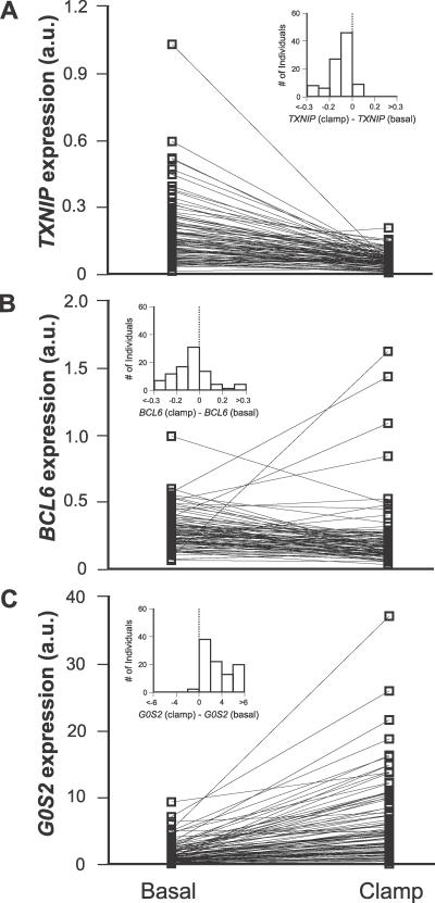
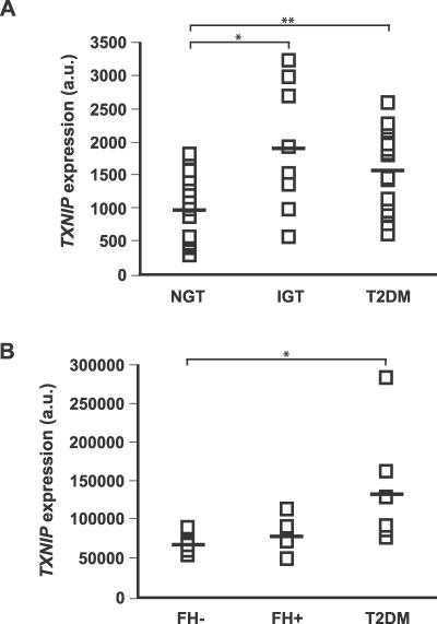
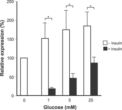
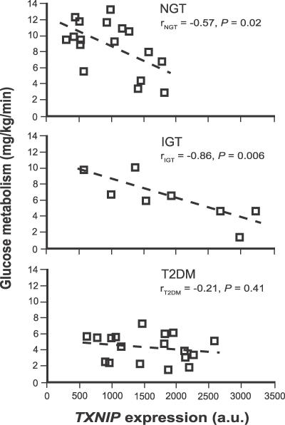
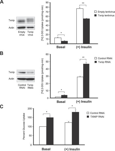
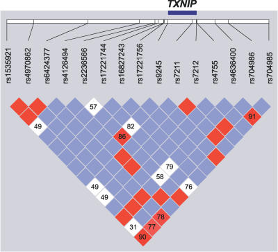
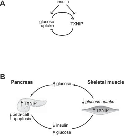
Methods and findings: We combined human insulin/glucose clamp physiological studies with genome-wide expression profiling to identify thioredoxin interacting protein (TXNIP) as a gene whose expression is powerfully suppressed by insulin yet stimulated by glucose. In healthy individuals, its expression was inversely correlated to total body measures of glucose uptake. Forced expression of TXNIP in cultured adipocytes significantly reduced glucose uptake, while silencing with RNA interference in adipocytes and in skeletal muscle enhanced glucose uptake, confirming that the gene product is also a regulator of glucose uptake. TXNIP expression is consistently elevated in the muscle of prediabetics and diabetics, although in a panel of 4,450 Scandinavian individuals, we found no evidence for association between common genetic variation in the TXNIP gene and T2DM.

Conclusions: TXNIP regulates both insulin-dependent and insulin-independent pathways of glucose uptake in human skeletal muscle. Combined with recent studies that have implicated TXNIP in pancreatic beta-cell glucose toxicity, our data suggest that TXNIP might play a key role in defective glucose homeostasis preceding overt T2DM.


Conflict of interest statement

Competing Interests: HT is employed at Novo Nordisk A/S, Denmark and owns a minor amount of employers stock in this Company. LCG is a member of the editorial board of PLoS Medicine.

Figures

Figure 1
Figure 1. Effects of Insulin on TXNIP, BCL6, and G0S2 Expression in Human Muscle In Vivo
mRNA levels were measured in skeletal muscle biopsies of healthy individuals before and after the hyperinsulinemic euglycemic clamp for (A) TXNIP (n = 96, p < 1 × 10⁻¹⁴), (B) BCL6 (n = 90, p < 1 × 10⁻⁵), and (C) G0S2 (n = 95, p < 1 × 10⁻¹⁶). Levels were measured using real-time PCR and normalized to cyclophilin A mRNA. Inset shows the distribution of expression changes over individuals. p-Values refer to the Wilcoxon signed rank statistic.
Figure 2
Figure 2. TXNIP Expression in Individuals with Diabetes or at Risk for Developing Diabetes
(A) TXNIP expression levels from individuals with NGT, IGT, or T2DM, using data from our previously published microarray study [18]. * p < 0.02; ** p < 0.01, Mann-Whitney U-test. (B) TXNIP expression levels from NGT individuals with family history of T2DM (FH+) or without (FH−), as well as individuals with T2DM, using data from a previously published microarray study [27]. * p < 0.03, Mann-Whitney U-test.
Figure 3
Figure 3. Effects of Glucose and Insulin on the Expression of TXNIP in Cultured Human Adipocytes
Human adipocytes were cultured in the presence (black bars) or absence (white bars) of 1 nM insulin at varying levels of glucose. Data are presented as mean ± standard deviation, normalized to the untreated control. * p < 0.05, Mann-Whitney U-test.
Figure 4
Figure 4. Insulin-Mediated Glucose Uptake Versus TXNIP Expression
This relationship is shown for individuals with NGT, IGT, and T2DM. Linear regression parameters are indicated for each of the groups. Data are from our previously published microarray study [18].
Figure 5
Figure 5. Genetic Manipulation of TXNIP Expression Affects Both Basal and Insulin-Stimulated Glucose Uptake in Cultured Adipocytes and in Primary Skeletal Muscle Myocytes
(A) Differentiated 3T3-L1 adipocytes cultured in 5.6 mM glucose were transduced with human TXNIP or empty virus particles and allowed to express the genes for 96 h. Cell lysates were immunoblotted using antibodies against TXNIP or actin. The exogenous viral human TXNIP protein is present as the more slowly migrating of the two bands. Glucose uptake with and without insulin was measured as described in Methods. Each data point represents an n = 4. * p < 0.02; ** p < 0.005. (B) Adipocytes cultured in 25 mM glucose were transfected with siRNA against TXNIP or with negative control siRNA and assayed 48 h posttransfection. Immunoblotting and glucose uptake are as above. n = 4 for each data point. * p < 0.02; ** p < 0.001. (C) Human skeletal muscle myoblasts obtained from biopsy were differentiated into myotubes in culture and transfected with siRNA against TXNIP or with negative control siRNA. Glucose uptake was measured at 72 h post-transfection with n = 4 for each data point. Each experiment was performed at least three times. * p < 0.01
Figure 6
Figure 6. Pattern of Genetic Variation at the TXNIP Locus
Linkage disequilibrium between SNPs was analyzed using Haploview [55], and D′ values were calculated with 95% confidence intervals. D′ plot for each square depicts the magnitude of linkage disequilibrium for a pair of markers, red indicates high D′ with high logarithm of odds (LOD) score, while white indicates low D′ with low LOD score, and blue indicates high D′ with low LOD score.
Figure 7
Figure 7. Model of TXNIP Regulation and Action and Its Potential Role in the Pathogenesis of T2DM
(A) Previous studies have shown that glucose induces the expression of TXNIP in a variety of cells and tissues. In this study we show that insulin suppresses the expression of TXNIP. Moreover, we have found that forced expression of TXNIP results in reduced glucose uptake, while inhibition of TXNIP enhances glucose uptake. These results suggest that TXNIP serves as a glucose- and insulin-sensitive homeostatic switch that regulates glucose uptake in the periphery. (B) Role of TXNIP in glucose toxicity in the β-cell and in impaired glucose uptake in the periphery. Insulin deficiency or hyperglycemia can increase TXNIP levels in muscle, resulting in impaired peripheral glucose uptake. The pancreatic β-cell is initially able to compensate by secreting more insulin, but eventually the β-cell compensation fails. The resulting hyperglycemia may then elevate pancreatic β-cell TXNIP expression, which can induce apoptosis [40]. The loss of β-cells, in turn, results in decreased insulin production that further exacerbates peripheral IGT. The vicious cycle would eventually spiral to T2DM.

References

    1. Kahn CR. Banting Lecture. Insulin action, diabetogenes, and the cause of type II diabetes. Diabetes. 1994;43:1066–1084. - PubMed
    1. Eriksson J, Franssila-Kallunki A, Ekstrand A, Saloranta C, Widen E, et al. Early metabolic defects in persons at increased risk for non-insulin-dependent diabetes mellitus. N Engl J Med. 1989;321:337–343. - PubMed
    1. Lyssenko V, Almgren P, Anevski D, Perfekt R, Lahti K, et al. Predictors of and longitudinal changes in insulin sensitivity and secretion preceding onset of type 2 diabetes. Diabetes. 2005;54:166–174. - PubMed
    1. Martin BC, Warram JH, Krolewski AS, Bergman RN, Soeldner JS, et al. Role of glucose and insulin resistance in development of type 2 diabetes mellitus: Results of a 25-year follow-up study. Lancet. 1992;340:925–929. - PubMed
    1. Groop L, Forsblom C, Lehtovirta M, Tuomi T, Karanko S, et al. Metabolic consequences of a family history of NIDDM (the Botnia study): Evidence for sex-specific parental effects. Diabetes. 1996;45:1585–1593. - PubMed

Publication types

MeSH terms

Associated data