. 2007 May 1;21(9):1086-97.
doi: 10.1101/gad.1535607.

Inhibition of U snRNP assembly by a virus-encoded proteinase

Affiliations

Inhibition of U snRNP assembly by a virus-encoded proteinase

Laura L Almstead et al. Genes Dev. .

Abstract

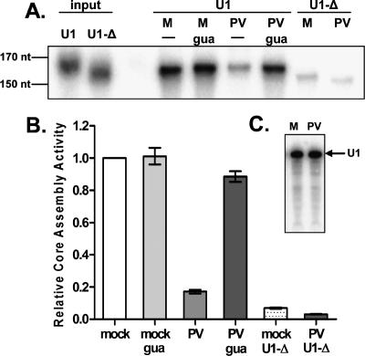
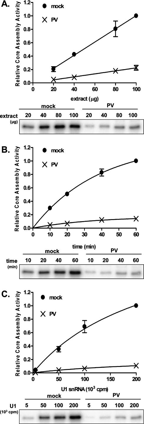
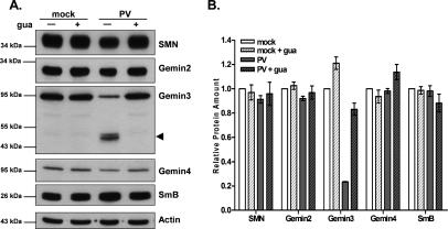
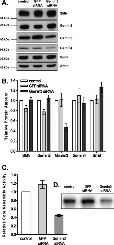
It has been proposed that defects in the assembly of spliceosomal uridine-rich small nuclear ribonucleoprotein (U snRNP) complexes could account for the death of motor neurons in spinal muscular atrophy (SMA). We discovered that infection of cultured cells with poliovirus results in the specific cleavage of the host factor Gemin3 by a virus-encoded proteinase, 2A(pro). Gemin3 is a component of the macromolecular SMN complex that mediates assembly of U snRNP complexes by aiding the heptameric oligomerization of Sm proteins onto U snRNAs. Using in vitro Sm core assembly assays, we found that lowering the intracellular amounts of Gemin3 by either poliovirus infection or small interfering RNA (siRNA)-mediated knockdown of Gemin3 resulted in reduced assembly of U snRNPs. Immunofluorescence analyses revealed a specific redistribution of Sm proteins from the nucleoplasm to the cytoplasmic periphery of the nucleus in poliovirus-infected cells. We propose that defects in U snRNP assembly may be shared features of SMA and poliomyelitis.


Figures

Figure 1.
Figure 1.
Sm core assembly in extracts from uninfected and poliovirus-infected cells. (A) Representative phosphorimage of U1 and U1-Δ snRNA core assembly reactions. Cells were infected with poliovirus (PV) or mock-infected (M), and assembly extracts were prepared 5 h post-infection. Addition of 2 mM guanidine-HCl (gua) was used to inhibit viral replication. Following assembly reactions, Sm cores were immunoprecipitated with an anti-Y12 antibody directed against epitopes of Sm proteins B′/B and D, and the associated snRNAs were resolved on 5% TBE-urea gels. The U1-Δ snRNA, containing a 9-nt deletion of the Sm core-binding site, served as a negative control. The input lanes on the left were loaded with 500 cpm of the indicated snRNAs. (B) Quantitation of three independent Sm core assembly experiments performed as described in A. Data were graphed as fractions of the U1 snRNA signal observed in mock-infected extracts. Error bars indicate the standard error. (C) Stability of U1 snRNAs during Sm core assembly reactions. Core assembly reactions were treated with proteinase K to disrupt RNA–protein interactions, and snRNAs were purified by phenol-chloroform extraction. Twenty percent of the material obtained from reactions containing mock-infected (M) or poliovirus-infected (PV) extracts was separated on 5% TBE-urea gels. A phosphorimage of the gel is shown. The position of the 164-nt U1 snRNA is indicated by an arrow.
Figure 2.
Figure 2.
Concentration dependence and kinetics of snRNP assembly in extracts from uninfected and infected cells. (A) U1 Sm core assembly as a function of extract concentration. (Top) Reactions were performed for 20 min at 30°C, and the amount of core-associated snRNA was plotted as a fraction of the core assembly observed in 100 μg of mock-infected extract. Data were fit to a first-order polynomial using GraphPad Prism to obtain a slope for each curve. Bottom panel shows a phosphorimage of a representative experiment. (B) Kinetics of U1 Sm core assembly. Standard Sm core assembly reactions were incubated for the indicated lengths of times and processed as in A. Data were plotted as the fraction of U1 snRNA signal observed at 60 min in mock-infected extracts. A one-phase exponential association equation (GraphPad Prism) was used to fit the data. Bottom panel shows a representative phosphorimage. (C) Sm core assembly as a function of U1 snRNA concentration. Increasing amounts of U1 snRNAs were used in standard core assembly reactions. Reactions were processed as in A. Top panel shows results plotted as the fraction of core assembly observed with 200,000 cpm U1 snRNA in mock-infected extracts. Data were fit to a one-site-binding hyperbola (GraphPad Prism). A representative phosphorimage is shown in the bottom panel. For each graph in A–C, error bars represent the standard error of three independent experiments. Error bars for some data points are too small to be visible.
Figure 3.
Figure 3.
Abundance and integrity of SMN complex and Sm core components in uninfected and infected cells. (A) Relative amounts of SMN complex components and Sm proteins in mock- and poliovirus-infected cells. Infection and treatment with guanidine-HCl (gua) were carried out as described in the legend for Figure 1A, and immunoblot analysis was performed using the indicated antibodies. Each lane contains 20 μg of total protein. The Gemin3 cleavage product is marked by an arrowhead. Molecular weight markers are shown on the left. (B) Quantitation of immunoblot analyses from experiments performed as described in A. For each antibody, the signal intensity was normalized to actin as a loading control, and the ratio obtained for mock-infected extracts was set equal to 1. Error bars represent the standard error of three independent experiments.
Figure 4.
Figure 4.
Effect of RNAi-mediated reduction of Gemin3 on Sm core assembly. (A) Immunoblot analysis of extracts prepared 48 h after transfection of siRNAs directed against GFP or Gemin3. Control extracts were obtained from cells treated with transfection reagent alone. Twenty micrograms of total protein from each sample were resolved by SDS-PAGE and blotted with the indicated antibodies. Molecular weight markers are shown on the left. (B) Quantitative analysis of the immunoblot analyses shown in A. For each antibody, the signal intensity was normalized to actin as a loading control, and the ratio obtained for control-treated extracts was set equal to 1. Error bars represent the standard error of three independent experiments. (C) Sm core assembly activity. Sm core assembly assays were performed using U1 snRNAs in extracts prepared from cells treated with transfection reagent alone (control), GFP siRNAs, or Gemin3 siRNAs. Assembly activity was plotted as the fraction of immunoprecipitated U1 snRNA observed in control extracts. Error bars represent the standard error of four independent experiments. (D) A representative phosphorimage of the Sm core assembly assays performed in C.
Figure 5.
Figure 5.
Effects of poliovirus infection on the colocalization of Sm proteins with coilin in nuclear foci. (A) Indirect immunofluorescence analysis of endogenous Sm and coilin proteins. HeLa cells plated on glass coverslips were fixed at 5 h post-mock or poliovirus infection and costained with anti-Y12 and anti-coilin antibodies. (Left panels) Differential interference contrast (DIC) images. (Middle panels) Hoechst-stained nuclei (blue). Representative maximum-intensity projections of deconvolved serial sections show the localization of Sm proteins (green) and coilin (red). (B) The number of colocalized foci observed in uninfected or poliovirus-infected cells were quantified. The graph represents the analysis of 100 cells for each condition. The p-value from χ2 analyses was <0.0001.
Figure 6.
Figure 6.
Cleavage activity of picornaviral protease 2Apro in vitro and in vivo. (A) Schematic representation of the Gemin3 protein sequence. The seven conserved helicase motifs (I–VI), the SMN interaction domain, and the region recognized by the Gemin3 antibody used in this study are indicated. The predicted 2A proteinase cleavage site is designated by an arrow. Amino acid residues are indicated below. (B) Immunoblot analysis of 2A proteinase cleavage reactions using antibodies against Gemin3 (top panel) or eIF4G (bottom panel). Lanes 1–3 contain samples from translation reactions in rabbit reticulocyte lysate with no RNA, wild-type Gemin3 RNA, or mutant Gemin3 RNA (Gemin3-G463E), respectively. Purified coxsackievirus B3 2A protease (2Apro) was added to the indicated samples, and cleavage reactions were incubated for 3 h at 37°C. For comparison, Gemin3 levels in total extracts prepared from cells that were infected for 5 h with poliovirus are shown in lane 8. An arrowhead marks the Gemin3 cleavage product. As a control, the blot was also incubated with antibodies directed against eIF4G to demonstrate that the recombinant protease was active as predicted. Due to the large molecular weight of eIF4G, the full-length protein transfers poorly and can be seen as a faint band marked as eIF4G. An asterisk shows the position of the cleaved eIF4G protein. (C) Cleavage of Gemin3 in cultured cells by transiently expressed polioviral 2A proteinase. Extracts were prepared from cells treated with transfection reagent alone (control), an expression vector lacking 2A coding sequences (pcDNA3.1), or a vector expressing the poliovirus 2A protease gene (2A-Flag) at 24 h post-transfection. Total protein extracts (20 μg) were separated using SDS-PAGE and blotted with an antibody directed against Gemin3. For comparison, 20 μg of total protein obtained from cells infected for 5 h with poliovirus was included. The Gemin3 cleavage product is indicated by an arrowhead. (D) Effects of poliovirus and rhinovirus 14 infection on Gemin3 cleavage. Extracts were prepared from mock-, rhinovirus 14-, and poliovirus-infected HeLa cells. Each lane contains 20 μg of total protein, and the blot was probed with the Gemin3 antibody. An arrowhead indicates the cleavage product.

References

    1. Baccon J., Pellizzoni L., Rappsilber J., Mann M., Dreyfuss G., Pellizzoni L., Rappsilber J., Mann M., Dreyfuss G., Rappsilber J., Mann M., Dreyfuss G., Mann M., Dreyfuss G., Dreyfuss G. Identification and characterization of Gemin7, a novel component of the survival of motor neuron complex. J. Biol. Chem. 2002;277:31957–31962. - PubMed
    1. Briese M., Esmaeili B., Sattelle D.B., Esmaeili B., Sattelle D.B., Sattelle D.B. Is spinal muscular atrophy the result of defects in motor neuron processes? Bioessays. 2005;27:946–957. - PubMed
    1. Buhler D., Raker V., Luhrmann R., Fischer U., Raker V., Luhrmann R., Fischer U., Luhrmann R., Fischer U., Fischer U. Essential role for the tudor domain of SMN in spliceosomal U snRNP assembly: Implications for spinal muscular atrophy. Hum. Mol. Genet. 1999;8:2351–2357. - PubMed
    1. Carissimi C., Saieva L., Baccon J., Chiarella P., Maiolica A., Sawyer A., Rappsilber J., Pellizzoni L., Saieva L., Baccon J., Chiarella P., Maiolica A., Sawyer A., Rappsilber J., Pellizzoni L., Baccon J., Chiarella P., Maiolica A., Sawyer A., Rappsilber J., Pellizzoni L., Chiarella P., Maiolica A., Sawyer A., Rappsilber J., Pellizzoni L., Maiolica A., Sawyer A., Rappsilber J., Pellizzoni L., Sawyer A., Rappsilber J., Pellizzoni L., Rappsilber J., Pellizzoni L., Pellizzoni L. Gemin8 is a novel component of the survival motor neuron complex and functions in small nuclear ribonucleoprotein assembly. J. Biol. Chem. 2006;281:8126–8134. - PubMed
    1. Carrel T.L., McWhorter M.L., Workman E., Zhang H., Wolstencroft E.C., Lorson C., Bassell G.J., Burghes A.H., Beattie C.E., McWhorter M.L., Workman E., Zhang H., Wolstencroft E.C., Lorson C., Bassell G.J., Burghes A.H., Beattie C.E., Workman E., Zhang H., Wolstencroft E.C., Lorson C., Bassell G.J., Burghes A.H., Beattie C.E., Zhang H., Wolstencroft E.C., Lorson C., Bassell G.J., Burghes A.H., Beattie C.E., Wolstencroft E.C., Lorson C., Bassell G.J., Burghes A.H., Beattie C.E., Lorson C., Bassell G.J., Burghes A.H., Beattie C.E., Bassell G.J., Burghes A.H., Beattie C.E., Burghes A.H., Beattie C.E., Beattie C.E. Survival motor neuron function in motor axons is independent of functions required for small nuclear ribonucleoprotein biogenesis. J. Neurosci. 2006;26:11014–11022. - PMC - PubMed

Publication types

MeSH terms