Case Reports
. 2007 May 2:2:12.
doi: 10.1186/1749-7221-2-12.

Platysma motor branch transfer in brachial plexus repair: report of the first case

Affiliations
Case Reports

Platysma motor branch transfer in brachial plexus repair: report of the first case

Jayme Augusto Bertelli. J Brachial Plex Peripher Nerve Inj. .

Abstract

Background: Nerve transfers are commonly employed in the treatment of brachial plexus injuries. We report the use of a new donor for transfer, the platysma motor branch.

Methods: A patient with complete avulsion of the brachial plexus and phrenic nerve paralysis had the suprascapular nerve neurotized by the accessory nerve, half of the hypoglossal nerve transferred to the musculocutaneous nerve, and the platysma motor branch connected to the medial pectoral nerve.

Results: The diameter of both the platysma motor branch and the medial pectoral nerve was around 2 mm. Eight years after surgery, the patient recovered 45 degrees of abduction. Elbow flexion and shoulder adduction were rated as M4, according to the BMC. There was no deficit after the use of the above-mentioned nerves for transfer. Volitional control was acquired for independent function of elbow flexion and shoulder adduction.

Conclusion: The use of the platysma motor branch seems promising. This nerve is expendable; its section led to no deficits, and the relearning of motor control was not complicated. Further anatomical and clinical studies would help to clarify and confirm the usefulness of the platysma motor branch as a donor for nerve transfer.


Figures

Figure 1
Figure 1
Schematic representation of the cervicomandibular branch of the facial nerve, its divisions and the surgical incision used to approach the platysma motor branch. (CFb) cervical branch of the facial nerve, which divides into the (Mb) mandibular branch and the (Cb) cervical branch. The Cb further divides into an (Ab) ascending branch, which is related to lower lip depression, and a (Db) descending branch, which innervates the lower portion of the platysma muscle. The Db is the branch used for transferring. (SM) submandibular gland.
Figure 2
Figure 2
Schematic representation of the surgical procedure to connect the (Pb) platysma motor branch to the (MP) medial pectoral nerve. A (SN) sural nerve graft was used to connect donor and recipient nerves.
Figure 3
Figure 3
Intraoperative view of the platysma motor branch. Scale bar = 2 mm
Figure 4
Figure 4
Intraoperative view of the medial pectoral nerve. Scale bar = 2 mm
Figure 5
Figure 5
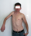
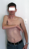
Results 8 years after surgery. The accessory nerve was connected to the suprascapular nerve, half of the hypoglossal nerve was grafted to the musculocutaneous nerve, and the platysma motor branch was transferred to the medial pectoral nerve. The patient recovered 45° of abduction and full elbow flexion, scoring M4. Shoulder adduction was restored with a M4 power. In 7, note shoulder adduction without concomitant elbow flexion. The independent control of these 2 functions is advantageous for the patient.
Figure 6
Figure 6
Results 8 years after surgery. The accessory nerve was connected to the suprascapular nerve, half of the hypoglossal nerve was grafted to the musculocutaneous nerve, and the platysma motor branch was transferred to the medial pectoral nerve. The patient recovered 45° of abduction and full elbow flexion, scoring M4. Shoulder adduction was restored with a M4 power. In 7, note shoulder adduction without concomitant elbow flexion. The independent control of these 2 functions is advantageous for the patient.
Figure 7
Figure 7
Results 8 years after surgery. The accessory nerve was connected to the suprascapular nerve, half of the hypoglossal nerve was grafted to the musculocutaneous nerve, and the platysma motor branch was transferred to the medial pectoral nerve. The patient recovered 45° of abduction and full elbow flexion, scoring M4. Shoulder adduction was restored with a M4 power. In 7, note shoulder adduction without concomitant elbow flexion. The independent control of these 2 functions is advantageous for the patient.

References

    1. Bertelli JA, Taleb M, Mira JC, Ghizoni MF. Functional recovery improvement is related to aberrant reinnervation trimming. A comparative study using fresh or predegenerated nerve grafts. Acta Neuropathol. 2006;2:601–609. doi: 10.1007/s00401-005-0005-0. - DOI - PubMed
    1. Chuang DCC. Neurotization procedures for brachial plexus injuries. Hand Clin. 1995;2:633–645. - PubMed
    1. Narakas AO. In: Microsurgery in orthopaedic practice. Leung PC, Gu YD, Ikuta Y, Narakas A, Landi A, Weiland AJ, editor. Singapore, World Scientific; 1995. Brachial plexus lesions; pp. 188–254.
    1. Bertelli JA, Ghizoni MF. Concepts of nerve regeneration and repair applied to brachial plexus reconstruction. Microsurgery. 2006;2:230–244. doi: 10.1002/micr.20234. - DOI - PubMed
    1. Cruveilhier J. Anatomie descriptive. Paris, Béchet Jeune, Tome IV; 1836. pp. 943–950.

Publication types