Comparative Study
. 2007 May 2:7:73.
doi: 10.1186/1471-2407-7-73.

Nectin-4 is a new histological and serological tumor associated marker for breast cancer

Affiliations
Comparative Study

Nectin-4 is a new histological and serological tumor associated marker for breast cancer

Stéphanie Fabre-Lafay et al. BMC Cancer. .

Abstract

Introduction: Breast cancer is a complex and heterogeneous disease at the molecular level. Evolution is difficult to predict according to classical histoclinical prognostic factors. Different studies highlight the importance of large-scale molecular expression analyses to improve taxonomy of breast cancer and prognostic classification. Identification of new molecular markers that refine this taxonomy and improve patient management is a priority in the field of breast cancer research.Nectins are cell adhesion molecules involved in the regulation of epithelial physiology. We present here Nectin-4/PVRL4 as a new histological and serological tumor associated marker for breast carcinoma.

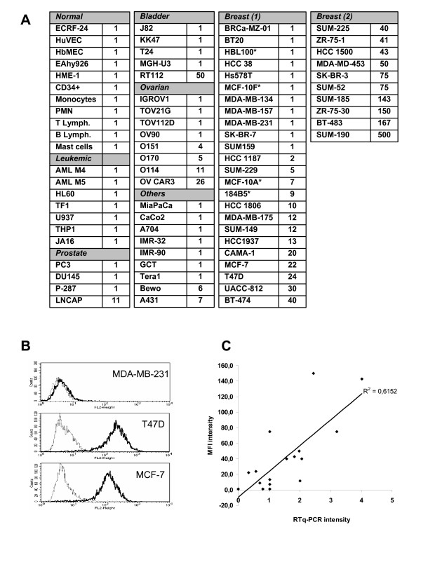
Methods: Expression of Nectin-4 protein was measured on a panel of 78 primary cells and cell lines from different origins and 57 breast tumors by FACS analysis and immunohistochemistry (IHC), respectively. mRNA expression was measured by quantitative PCR. Serum Nectin-4 was detected by ELISA and compared with CEA and CA15.3 markers, on panels of 45 sera from healthy donors, 53 sera from patients with non-metastatic breast carcinoma (MBC) at diagnosis, and 182 sera from patients with MBC. Distribution of histological/serological molecular markers and histoclinical parameters were compared using the standard Chi-2 test.

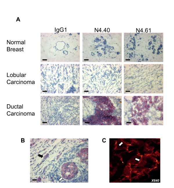
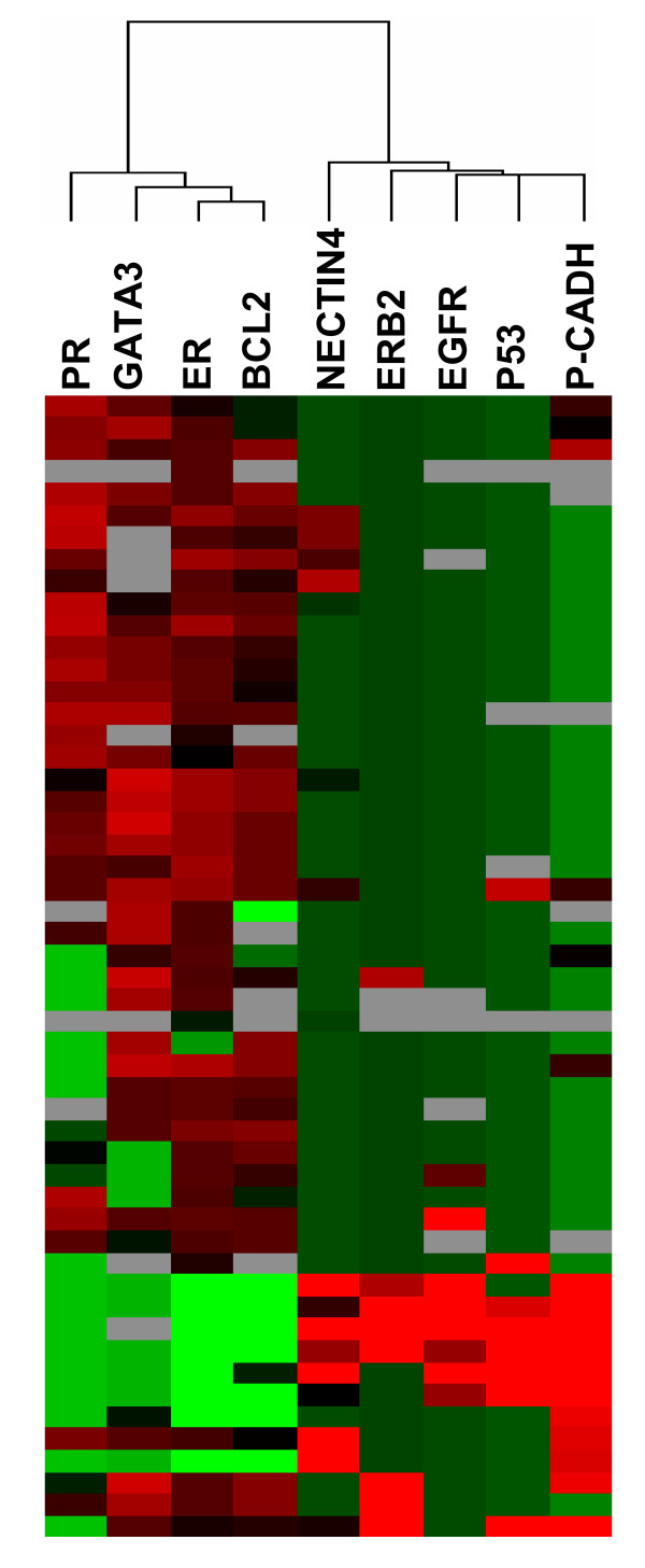
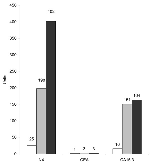
Results: Nectin-4 was not detected in normal breast epithelium. By contrast, Nectin-4 was expressed in 61% of ductal breast carcinoma vs 6% in lobular type. Expression of Nectin-4 strongly correlated with the basal-like markers EGFR, P53, and P-cadherin, and negatively correlated with the luminal-like markers ER, PR and GATA3. All but one ER/PR-negative tumors expressed Nectin-4. The detection of Nectin-4 in serum improves the follow-up of patients with MBC: the association CEA/CA15.3/Nectin-4 allowed to monitor 74% of these patients compared to 67% with the association CEA/CA15.3. Serum Nectin-4 is a marker of disease progression, and levels correlate with the number of metastases (P = 0.038). Serum Nectin-4 is also a marker of therapeutic efficiency and correlates, in 90% of cases, with clinical evolution.

Conclusion: Nectin-4 is a new tumor-associated antigen for breast carcinoma. Nectin-4 is a new bio-marker whose use could help refine breast cancer taxonomy and improve patients' follow-up. Nectin-4 emerges as a potential target for breast cancer immunotherapy.


Figures

Figure 1
Figure 1
Analysis of Nectin-4 expression in normal and tumor cells. A: Cell surface expression of Nectin-4 was assessed by FACS analysis using the N4.61 mAb. The level of cell surface expressed Nectin-4 (L) was calculated in arbitrary units based on fluorescence intensity: L = MFI N4.61/MFI control IgG1. Similar results were obtained with the N4.40 mAb. L = 1 corresponds to negative cell lines, 2 < L < 15: low expression, 16 < L < 50: intermediate expression, L > 50 high expression. These results are representative of at least three independant experiments. *, non cancerous breast cell lines. B: Nectin-4 expression level on breast tumor cell lines was monitored by FACS analysis using the anti-Nectin-4 N4.61 mAb (black line) and compared with a mouse irrelevant IgG1 (gray line). Examples of a negative cell line (MDA-MB-231) and two intermediate expressing cell lines (T47D and MCF-7). C: Correlation between Nectin-4 cell surface expression and Nectin-4 transcriptional expression. Quantitative PCR was performed on 17 breast tumor cell lines. Values were calculated as described in material and methods. The results are representative of at least two experiments.
Figure 2
Figure 2
Nectin-4 expression in breast carcinoma. A: Normal breast epithelium does not express Nectin-4. No expression was detected in luminal, myoepithelial and stromal cells. Nectin-4 expression was found prominently in ductal carcinoma. Bar 50 μm. Magnification: × 200. B: Both invasive (black arrow) and in situ (white arrow) components of ductal carcinoma are immunostained by Nectin-4. C: Immunofluorescence analysis revealed that Nectin-4 expression is cytoplasmic (arrowhead) and junctional (arrow). Similar results were obtained with both mAbs Magnification: × 640.
Figure 3
Figure 3
Nectin-4 expression correlates with basal-like breast markers. Graphical representation of hierarchical clustering results based on expression profiles of 9 proteins in 52 early breast cancer samples measured by immunohistochemistry. Rows: proteins; columns: samples. Protein expression scores are depicted according to a color scale: red, positive staining; green, negative staining; grey, missing data. Dendrograms represent overall similarities in protein expression profiles. Two clusters of proteins are described. A group of proteins, including Nectin-4, which present high expression profile similarities with basal breast markers. A group of proteins, including luminal markers, which negatively correlate with Nectin-4 expression and basal-like markers.
Figure 4
Figure 4
Detection of Nectin-4 in serum from patients with MBC. Serum Nectin-4 was detected by ELISA using a pair of anti-Nectin-4 mAbs. A: Percentage of Nectin-4 positive sera in healthy donors (n = 45), and in patients with non-metastatic breast tumors compared with CEA and CA15.3 levels (n = 53). B: Percentage of sera positive for Nectin-4, CEA and CA15.3 and combinations of markers. The association of Nectin-4 with CEA and CA15.3 increases the percentage of patients detected. White bars; patients with metachronous MBC. Grey bars; patients with synchronous MBC. Black bars; percentage including both groups. C: ROC curves were calculated to estimate the accuracy of the association of these markers in breast cancer diagnosis.
Figure 5
Figure 5
Serum Nectin-4 and disease progression. Nectin-4, CEA and CA15.3 serum levels were determined during disease progression of a patient with a ductal carcinoma. The three markers were not detected at the time of diagnosis (white bar). During the progression of the disease, Nectin-4 serum levels increased to reach 402 pM. Grey, detection of pulmonary metastasis 32 months post-diagnosis; black, detection of brain metastasis 39 months post-diagnosis. Ordinates represent pM for Nectin-4 and International Units for CEA and CA15.3.
Figure 6
Figure 6
Serum Nectin-4 and therapeutic efficiency. Nectin-4, CEA and CA15.3 levels were determined in the serum of patients at the time of metastasis diagnosis and after therapy. Results are represented by arrows that symbolize increased, apparition, decreased, extinction, or unchanged (formula image
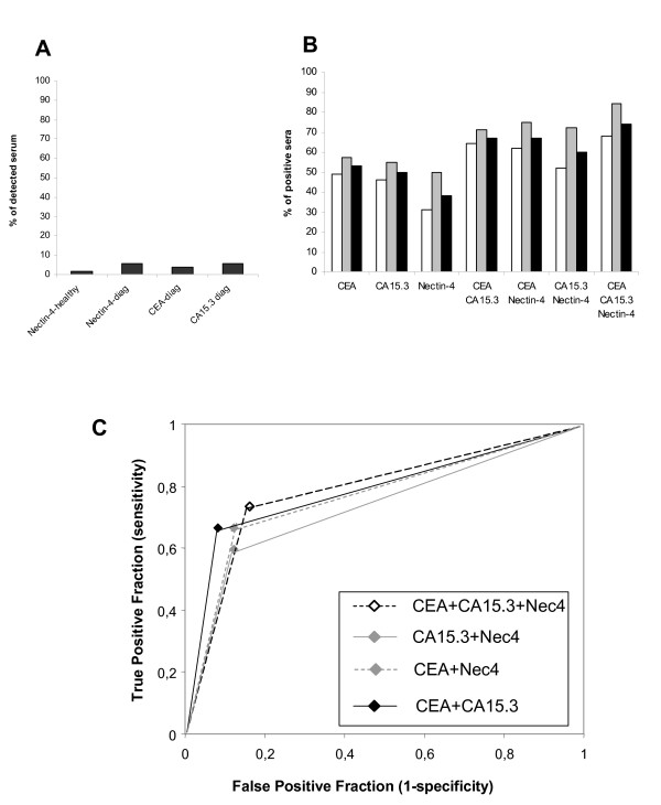
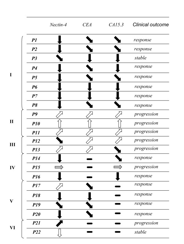
) levels of Nectin-4, CEA and CA15.3. Patient outcome was evaluated according to clinical and histoclinical criteria.

References

    1. Baselga J, Norton L. Focus on breast cancer. Cancer Cell. 2002;1:319–322. doi: 10.1016/S1535-6108(02)00066-1. - DOI - PubMed
    1. Ludwig JA, Weinstein JN. Biomarkers in cancer staging, prognosis and treatment selection. Nat Rev Cancer. 2005;5:845–856. doi: 10.1038/nrc1739. - DOI - PubMed
    1. Perou CM, Sorlie T, Eisen MB, van de Rijn M, Jeffrey SS, Rees CA, Pollack JR, Ross DT, Johnsen H, Akslen LA, Fluge O, Pergamenschikov A, Williams C, Zhu SX, Lonning PE, Borresen-Dale AL, Brown PO, Botstein D. Molecular portraits of human breast tumours. Nature. 2000;406:747–752. doi: 10.1038/35021093. - DOI - PubMed
    1. Sorlie T, Tibshirani R, Parker J, Hastie T, Marron JS, Nobel A, Deng S, Johnsen H, Pesich R, Geisler S, Demeter J, Perou CM, Lonning PE, Brown PO, Borresen-Dale AL, Botstein D. Repeated observation of breast tumor subtypes in independent gene expression data sets. Proc Natl Acad Sci U S A. 2003;100:8418–8423. doi: 10.1073/pnas.0932692100. - DOI - PMC - PubMed
    1. Bertucci F, Finetti P, Rougemont J, Charafe-Jauffret E, Cervera N, Tarpin C, Nguyen C, Xerri L, Houlgatte R, Jacquemier J, Viens P, Birnbaum D. Gene expression profiling identifies molecular subtypes of inflammatory breast cancer. Cancer Res. 2005;65:2170–2178. doi: 10.1158/0008-5472.CAN-04-4115. - DOI - PubMed

Publication types