Review
. 2007 May 2:2:9.
doi: 10.1186/1749-8104-2-9.

Drosophila as a genetic and cellular model for studies on axonal growth

Affiliations
Review

Drosophila as a genetic and cellular model for studies on axonal growth

Natalia Sánchez-Soriano et al. Neural Dev. .

Abstract

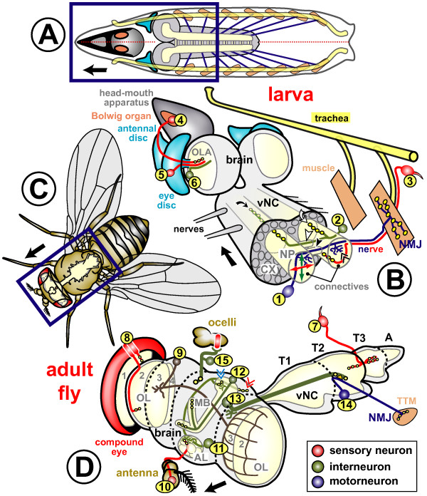
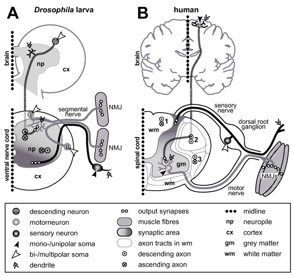
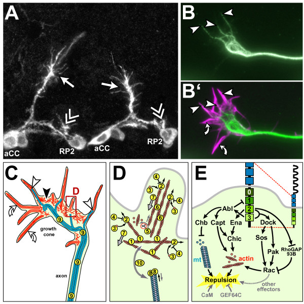
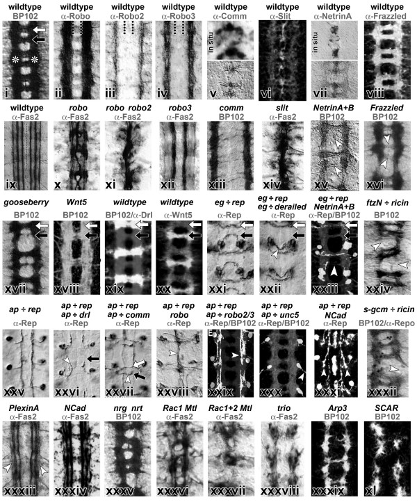
One of the most fascinating processes during nervous system development is the establishment of stereotypic neuronal networks. An essential step in this process is the outgrowth and precise navigation (pathfinding) of axons and dendrites towards their synaptic partner cells. This phenomenon was first described more than a century ago and, over the past decades, increasing insights have been gained into the cellular and molecular mechanisms regulating neuronal growth and navigation. Progress in this area has been greatly assisted by the use of simple and genetically tractable invertebrate model systems, such as the fruit fly Drosophila melanogaster. This review is dedicated to Drosophila as a genetic and cellular model to study axonal growth and demonstrates how it can and has been used for this research. We describe the various cellular systems of Drosophila used for such studies, insights into axonal growth cones and their cytoskeletal dynamics, and summarise identified molecular signalling pathways required for growth cone navigation, with particular focus on pathfinding decisions in the ventral nerve cord of Drosophila embryos. These Drosophila-specific aspects are viewed in the general context of our current knowledge about neuronal growth.


Figures

Figure 1
Figure 1
Neurons used for studies on neuronal growth at different stages of Drosophila development. (a,c) Horizontal views of a Drosophila larva and adult fly, respectively, illustrating the position of the CNS (grey and cream) in relation to other body structures. (b,d) Three-dimensional extracts from the areas boxed in dark blue in (a,c), respectively. The cell body area of the CNS (cortex (CX)) is shown in light grey, and the neuritic/synaptic area (neuropile (NP)) in cream (only relevant neuropile structures are shown in (b,d)). Black arrows point anterior, morphological structures are annotated in colour code, and neuronal classes are explained in the box at bottom right. The various model neurons are marked with numbers in yellow circles, explained below. Many neurons of the larval trunk can be studied from their birth in the embryo through to the mature synaptic stage. Amongst these, motorneurons (1) project towards the dorsal zone of ipsilateral or ipsi- and contralateral connectives (where they form dendrites; double chevron), from where they enter specific branches of peripheral nerves leading towards their target muscles, on which they form neuromuscular junctions (NMJ; yellow circles represent chemical synapses). Projections of larval interneurons (2) are restricted to the neuropile. Sensory neurons of the trunk (3) project along tracheal branches and motoraxons towards the ventral nerve cord (vNC) where they innervate the ventral domain of connectives [192,195,196,198-200,236]. Sensory neurons in the embryonic trunk have been used, for example, to study the actin-microtubule linker molecule Short stop, signalling through Robo or Notch receptors, or the spatial arrangement of axons in the neuropile [197,202,203,255]. Projections of neurons 1, 2 and 3 in the neuropile of the ventral nerve cord can be classified with respect to their anteroposterior extension within the segment (white curved arrow) or across segments (black curved arrow), their dorsoventral and mediolateral position in connectives (green and red double arrows, respectively), their ipsilateral (neuron 3) versus contralateral (neurons 1 and 2) nature, and their projection through anterior (white arrowhead) versus posterior commissure (black arrowhead; see details in 'Signalling mechanisms involved in axonal pathfinding in Drosophila' above). In the embryonic/larval head region (4), the Bolwig organ has been used for studies of neuronal growth. It contains somata of 12 photoreceptor cells [306], the axons of which form the Bolwig nerve projecting over the antennal and eye discs via the optic stalk into the optic lobe anlage (OLA) [26,307]. The Bolwig nerve is joined by successively outgrowing waves of axons of photoreceptor neurons (5), which are specified in the eye disc during larval and pupal stages. The optic lobe pioneer neuron (6), a projection neuron of embryonic origin, seems to be used as a guide within the OLA by the Bolwig nerve and photoreceptor axons [308,309]. Sensory neurons of the adult trunk (7) develop de novo during larval and pupal stages (with a few exceptions) [310] and terminate in the vNC neuropile (T1-3 and A indicate the three thoracic and fused abdominal segments). They can be analysed from the time of birth through to the fully differentiated stage [311,312], and have been used to study features, such as segment-specific growth regulation (homeotic genes), or the influence of adhesive interactions (Dscam), axonal transport (cut up, the dynein light chain) or of size alterations (gigas) on neuronal growth behaviour [311,313-315]. Photoreceptor cells in the adult compound eye (8) form a precise retinotopic map in the optic lobe (OL: grey 1, lamina; 2, medulla; 3, lobula; 4, lobula plate) established during larval (see neuron 5) and pupal stages, and the genetic mechanisms regulating these precise growth decisions are beginning to be unravelled [316-318]. Interneurons postsynaptic to photoreceptor neurons are well described [317,319] but seem not to have been used for studies of growth mechanisms so far, with the exception of a group of 20–30 dorsal cluster neurons (9; targeted by atoGal4-14A), which form dendrites in the ipsilateral optic lobe and project through the dorsal commissure to innervate the contralateral lobula and medulla [320-322]. Olfactory neurons in the third antennal segment (10) and the maxillary palp (not shown) project from the antenna into the antennal lobe (AL) where they terminate in specific glomeruli in a reproducible pattern correlating with the class of odorant receptor they express; the genetic regulation of this growth behaviour is under investigation [39]. The major output from the AL is constituted by projection neurons (11), which are postsynaptic to olfactory neurons and innervate the lateral horn (red double chevron) and the calyx (blue double chevron), a dorsal structure of the mushroom bodies (MB) [39]. The mushroom bodies are the brain structures responsible for olfactory learning in Drosophila [323,324], and its intrinsic interneurons (Kenyon cells (12)) project through the calyx and pedunculus where many of them bifurcate to project into the vertical α/α'-and the horizontal β/β'/γ-lobes, simultaneously [325]. The large giant fibre neuron (13) connects the optic system via a large diameter axon with motorneurons in the second thoracic segment (14), innervating the tergotrochanteral muscle (TTM; responsible for the visually induced jump escape response) via chemical and electrical (orange triangle) synapses [326]. Giant fibre axons grow out during late larval/pupal stages and have been used to study growth regulatory mechanism, such as the influence of Rho-like GTPases or the role of the E2 ubiquitin ligase Bendless [326]. Ocellar photoreceptor neurons do not send out their own axons but are connected to the brain via large interneurons, the cell bodies of which are located in the brain originally, but migrate into the periphery during pupal development (15). The pathfinding of these interneurons depends on a set of short-lived pioneer neurons that, in turn, require the extracellular matrix molecule laminin, the transmembrane receptor neurotactin and its ligand Amalgam for proper outgrowth [21,239,241]. Further potentially attractive models for studies of neuronal growth that are not shown here are auditory sensory neurons [327], and axonal fascicles in the ventral nerve cord of late Drosophila larvae representing paused interneurons of the future adult CNS (not shown) [209].
Figure 2
Figure 2
Comparing principal features of neuronal organisation and growth in Drosophila (larva) and vertebrates (human). (a) Saggital section (one body half; dotted line is midline) through the larval brain and ventral nerve cord (compare Figure 1a,b). (b) Saggital section through the adult human brain and one half of the spinal cord. Symbols are explained in the box below. Whereas axons of unipolar inter- and motorneurons in Drosophila have to grow into the synaptic area where they form dendrites, comparable neurons in vertebrates are multipolar and locate themselves in the synaptic area. All Drosophila motorneurons locate their dendrites in the dorsal neuropile, regardless of their soma position (see 'i' versus 'ii') [236]. Vice versa, sensory somata in Drosophila are located next to their dendrites, whereas cell bodies of most sensory neurons in vertebrates are grouped together in the dorsal root ganglia. Sensory output (dark grey) and motor input areas (bright grey) are inverted in both phyla, which might be explained through a general dorsoventral body axis inversion between vertebrates and arthropods [328], that is, not represent an organisational difference between their CNS. Ascending/descending axons in Drosophila are non-myelinated and project through the synaptic area (compare neurons 2 and 13 in Figure 1) where they take on characteristic positions [236,329]. In vertebrates, ascending/descending axons are myelinated and positioned outside the synaptic area, grouping into characteristic tracts in defined positions of the white matter; examples named here: fasciculus cuneatus (1), tractus corticospinalis lateralis (2; pyramidal tract; only descending), and tractus spinothalamicus lateralis (3).
Figure 3
Figure 3
Drosophila growth cones and the (potential) factors regulating their cytoskeletal dynamics. (a) Growth cones of aCC (arrows) and RP2 motorneurons (double chevrons; cell bodies named) in two consecutive segments of the trunk of a Drosophila embryo, stained with a cell-specifically expressed membrane marker. (b,b') Cultured Drosophila growth cone stained for microtubules (green) and filamentous actin (magenta); some filopodia lack microtubules (curved arrows), whereas others are deeply invaded (arrow heads indicate microtubule tips). (c) Schematic representation of the cytoskeletal organisation in Drosophila growth cones as extrapolated from work on growth cones in other species (detailed in the section 'Principal structure and function of growth cones'): veil-like lamellipodia (black arrowhead) contain mesh-like networks of actin filaments (randomly oriented red lines), whereas pointed filopodia (white arrowhead) contain bundled actin filaments (parallel red lines); microtubules (blue lines) are bundled in the axon, but single splayed microtubules extend into the periphery of the growth cone (curved white arrows indicate splayed microtubule tips), reaching into filopodia, as was similarly reported for growth cones of other species or migrating cells [63,330]. (d) Details of the boxed area in (c); circled numbers correlate with the numbers in Table 1 and represent the following molecular activities: 1, actin filament nucleation by Arp2/3 (which subsequently stays with the pointed ends); 2, actin filament nucleation and elongation by formins (which stay with barbed ends); 3, actin monomer binding; 4, barbed-end capping; 5, pointed end-depolymerisation/severing; 6, actin filament bundling; 7, retrograde flow of actin cytoskeleton; 8, microtubule plus end binding; 9, microtubule stabilising; 10, actin-microtubule linkage. Black straight arrows indicate growth of actin filaments or microtubules, grey straight arrows shrinkage, black curved arrows addition of actin monomers, grey curved arrows removal of actin monomers or filamentous fragments, hatched arrows indicate direction of retrograde actin flow, and the grey dashed curved double arrow linkage of actin and microtubules. (e) Current view of the effectors downstream of the Slit receptor Robo mediating repulsion from the midline of the ventral nerve cord. Robo (top right) habours five immunoglobulin domains (half elipses) and three fibronectin type III domains (blue boxes) extracellularly, and four conserved cytoplasmic (CC) domains (light to dark green) intracellularly. Robo induces growth cone repulsion by controlling cytoskeletal dynamics via either Abelson kinase (Abl) and Enabled (Ena), or Rac activity. Ena binds at CC2 and acts most likely through Chickadee/Profilin on actin dynamics. Abl binding to Robo at CC3 influences actin dynamics via Capulet and microtubule dynamics via the +TIP protein Chromosome Bows (Chb/Orbit/MAST). Simultaneously, Abl phosphorylates CC1 to antagonise Robo function. The regulation of Rac activity through Robo occurs through CC2/3 recruitment of the SH3-SH2 adaptor molecule Dreadlocks (Dock) which, in turn, activates Rac through both Pak and the GEF Sos. In parallel, active Robo can influence Rac activity via the binding of RhoGAP93B (vilse/CrGAP) to CC2, but it remains unclear whether RhoGAP93B is positively or negatively regulated by Robo. Paradoxically, both decrease and increase of Rac activation levels can cause midline crossing, suggesting that: Rac might influence other effectors to cause repulsion; a precise Rac activation level is required to mediate Slit-induced repulsion; or a sequential modification of Rac in response to Robo activation has to occur, such as an initial role to prevent extension towards the source of the repellent and another role to encourage extension away from the Slit source. Calmodulin and GEF64C have additionally been identified as modifiers of Robo activity, although it is not clear yet how they influence Robo signalling (calmodulin possibly through Chic).
Figure 4
Figure 4
Representative embryonic mutant phenotypes of axonal projections in the ventral nerve cord. Images of ventral nerve cords in horizontal view (dorsal up) of embryos that are wild type, mutant or display targeted expression of genes in subsets of neurons. Genotypes are indicated in black at the top, antibody stainings in grey (abbreviations: Robo, Roundabout; Comm, Commissureless; Fas2, Fasciclin 2; Drl, Derailed; Rep, reporter gene; eg, eagle; ftz, fushi tarazu; ap, apterous; unc5, uncoordinated 5; NCad, N-Cadherin; gcm, glia cells missing; nrg, neuroglian; nrt, neurotactin; "X ÷ Y", expression of gene Y driven by the promoter of gene X). BP102 antiserum labels the complete neuropile (i), consisting of two connectives (asterisks) and, per segment, an anterior (white arrow) and posterior (black arrow) commissure. At stage 16 Fasciclin 2 labels three prominent longitudinal fascicles per connective (ix). (ii-viii) Expression patterns of genes involved in midline crossing and, below (x-xvi), respective loss-of-function phenotypes (see text for details). (xvii-xxii) Regulation of growth through the anterior versus posterior commissure, with loss of posterior (xvii) or anterior (xviii) commissure, specific expression of Derailed (xix) in anterior and its ligand Wnt (xx) in posterior commissure, and shift of posterior commissure neurons (eagle-Gal4; xxi) to the anterior commissure (white arrow head in xxii) upon Derailed expression in their axons. (xxiii) Same eagle-Gal4 neurons partially lack commissural projections in netrin A+B mutant background (compare white arrowhead in (xv)). (xxv-xxxi) Detailed phenotypic studies using identified apterous-Gal4 neurons, which project transversely to the medial connectives where they form a longitudinal fascicle (xxv); as indicated by white arrows, they stall prematurely upon Derailed expression (xxvi), project across the midline upon Comm expression (xxvii), collapse towards the midline in robo mutant background (compare x), shift to lateral positions upon Robo2+3 expression (xxix), project out of the CNS upon Unc5 expression (xxx), or turn prematurely from transverse into longitudinal direction in N-cadherin mutant background (xxxi). (xxiv, xxxii). Connectives are affected upon ablation of longitudinal pioneer neurons (xxiv) or longitudinal glia cells (xxxii). (xxxiii-xl) Various neuropile phenotypes in embryos mutant for transmembrane molecules (xxxiii-xxxv) or factors involved in cytoskeletal regulation (xxxvi-xl). Images were taken, with permission, from [241] (i, xxxv), [253] (ii-iv, xii, xxix), [207] (v, top), [331] (v, bottom), [259] (vi), [266] (vii, xv), [225] (viii), [244] (ix, xxxiii), [271] (x, xi, xiv), [23] (xiii, xvii), [212] (xvi, xxx), [263] (xviii), [208] (xix, xxi, xxii, xxv-xxvii), [332] (xx), [267] (xxiii), [219] (xxiv), [277] (xxviii), [226] (xxxi, xxxiv), [223] (xxxii), [132] (xxxvi-xxxviii), [95] (xxxix, xxxx). Images were modified to grayscale and adapted to size.
Figure 5
Figure 5
Axonal pathfinding and fasciculation behaviour in the embryonic ventral nerve cord. (a) In the ventral nerve cord of stage 13/14 embryos, growth cones of identified neurons (aCC, curved arrow; pCC, arrow; RP2, arrow head) navigate in stereotypic positions (stippled line, midline; asterisk, somata of aCC and pCC). (b) Schematic representations of early growing neurons vMP2, dMP2, MP1, aCC and pCC (colour coded): at stage 13, their axons are partly guided by glia cells (grey circles) and Netrin A and B (light green) bound to lateral fields of Frazzled expression (wave pattern); MP1s and dMP2s grow jointly posteriorward, whereas vMP2s and pCCs fasciculate and grow together anteriorward until all four neurons contact one another midway between adjacent neuromeres and establish a single longitudinal fascicle (stage 13) that splits (stage 14), re-fasciculates (stage 15) and splits again (stage 16), partly mediated by glia cells (pCCs, dMP2s, vMP2s form a common fascicle close to the midline, MP1s a distinct axon tract further lateral; grey stippled line represents the lateral Fas2 fascicle of unknown identity; compare Figure 4, ix). (c,d) The neuroblast lineage NB1-2 [190] illustrates the stereotypic pathfinding choices of individual neurons (curved arrow, ipsilateral longitudinal path; AC/PC, anterior/posterior commissure; arrow, medial contralateral longitudinal; double chevron, lateral contralateral longitudinal; arrow head, soma of identified TB neuron; CX, cortex; NP, neuropile). (e) Regulation of midline crossing and mediolateral longitudinal path choice: ipsilateral neurons don't express Commissureless (Comm), and their combinatorial Robo receptor code determines the mediolateral positioning of their axons; contralateral neurons express Comm (black T), thus preventing transport of Robo receptors to the growth cone (curved red arrow); subsequent downregulation of Comm activity permits the Robo-mediated fascicle choice. (f) Choice of anterior versus posterior commissure during midline crossing is partly determined by posterior expression of Wnt5 (Figure 4, xx), which repels growth cones of Derailed expressing neurons. (c,d) Kindly provided by Janina Seibert, Christoph Rickert and Gerd Technau; (b) redrawn from Hidalgo and Booth [223].

References

    1. Ramón y Cajal S. Histology of the Nervous System of Man and Vertebrates (English translation by Swanson N, Swanson LW from 1995) New York Oxford: Oxford University Press; 1909. p. 1911.
    1. Tessier-Lavigne M, Goodman CS. The molecular biology of axon guidance. Science. 1996;274:1123–1133. - PubMed
    1. Araujo SJ, Tear G. Axon guidance mechanisms and molecules: lessons from invertebrates. Nat Rev Neurosci. 2003;4:910–922. - PubMed
    1. Bate CM. Pioneer neurones in an insect embryo. Nature. 1976;260:54–56. - PubMed
    1. Harrison RG. The outgrowth of the nerve fiber as a mode of protoplasmic movement. J Exp Zool. 1910;9:787–846. - PubMed

Publication types

MeSH terms

LinkOut - more resources