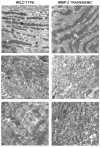
Transgenic MMP-2 expression induces latent cardiac mitochondrial dysfunction
- PMID: 17475219
- PMCID: PMC3423089
- DOI: 10.1016/j.bbrc.2007.04.094
Transgenic MMP-2 expression induces latent cardiac mitochondrial dysfunction
Abstract
Matrix metalloproteinases (MMPs) are central to the development and progression of dysfunctional ventricular remodeling after tissue injury. We studied 6 month old heterozygous mice with cardiac-specific transgenic expression of active MMP-2 (MMP-2 Tg). MMP-2 Tg hearts showed no substantial gross alteration of cardiac phenotype compared to age-matched wild-type littermates. However, buffer perfused MMP-2 Tg hearts subjected to 30 min of global ischemia followed by 30 min of reperfusion had a larger infarct size and greater depression in contractile performance compared to wild-type hearts. Importantly, cardioprotection mediated by ischemic preconditioning (IPC) was completely abolished in MMP-2 Tg hearts, as shown by abnormalities in mitochondrial ultrastructure and impaired respiration, increased lipid peroxidation, cell necrosis and persistently reduced recovery of contractile performance during post-ischemic reperfusion. We conclude that MMP-2 functions not only as a proteolytic enzyme but also as a previously unrecognized active negative regulator of mitochondrial function during superimposed oxidative stress.
Figures


Similar articles
-
Intracellular regulation of matrix metalloproteinase-2 activity: new strategies in treatment and protection of heart subjected to oxidative stress.Scientifica (Cairo). 2013;2013:130451. doi: 10.1155/2013/130451. Epub 2013 Dec 24. Scientifica (Cairo). 2013. PMID: 24455428 Free PMC article. Review.
-
Efficacy of ischaemic preconditioning in the eNOS overexpressed working mouse heart model.Eur J Pharmacol. 2007 Feb 5;556(1-3):115-20. doi: 10.1016/j.ejphar.2006.11.004. Epub 2006 Nov 10. Eur J Pharmacol. 2007. PMID: 17157294
-
Myocardial recovery from ischemia-reperfusion is compromised in the absence of tissue inhibitor of metalloproteinase 4.Circ Heart Fail. 2014 Jul;7(4):652-62. doi: 10.1161/CIRCHEARTFAILURE.114.001113. Epub 2014 May 19. Circ Heart Fail. 2014. PMID: 24842912
-
Cardiac-specific overexpression of fibroblast growth factor-2 protects against myocardial dysfunction and infarction in a murine model of low-flow ischemia.Circulation. 2003 Dec 23;108(25):3140-8. doi: 10.1161/01.CIR.0000105723.91637.1C. Epub 2003 Dec 1. Circulation. 2003. PMID: 14656920
-
Targeting MMP-2 to treat ischemic heart injury.Basic Res Cardiol. 2014 Jul;109(4):424. doi: 10.1007/s00395-014-0424-y. Epub 2014 Jul 2. Basic Res Cardiol. 2014. PMID: 24986221 Review.
Cited by
-
Transgenic expression of matrix metalloproteinase-2 induces coronary artery ectasia.Int J Exp Pathol. 2011 Feb;92(1):50-6. doi: 10.1111/j.1365-2613.2010.00744.x. Epub 2010 Oct 29. Int J Exp Pathol. 2011. PMID: 21039989 Free PMC article.
-
A novel intracellular isoform of matrix metalloproteinase-2 induced by oxidative stress activates innate immunity.PLoS One. 2012;7(4):e34177. doi: 10.1371/journal.pone.0034177. Epub 2012 Apr 3. PLoS One. 2012. PMID: 22509276 Free PMC article.
-
Intracellular regulation of matrix metalloproteinase-2 activity: new strategies in treatment and protection of heart subjected to oxidative stress.Scientifica (Cairo). 2013;2013:130451. doi: 10.1155/2013/130451. Epub 2013 Dec 24. Scientifica (Cairo). 2013. PMID: 24455428 Free PMC article. Review.
-
A N-terminal truncated intracellular isoform of matrix metalloproteinase-2 impairs contractility of mouse myocardium.Front Physiol. 2014 Sep 25;5:363. doi: 10.3389/fphys.2014.00363. eCollection 2014. Front Physiol. 2014. PMID: 25309453 Free PMC article.
-
Immunosuppression With FTY720 Reverses Cardiac Dysfunction in Hypomorphic ApoE Mice Deficient in SR-BI Expression That Survive Myocardial Infarction Caused by Coronary Atherosclerosis.J Cardiovasc Pharmacol. 2016 Jan;67(1):47-56. doi: 10.1097/FJC.0000000000000312. J Cardiovasc Pharmacol. 2016. PMID: 26322923 Free PMC article.
References
-
- Nagase H, Visse R, Murphy G. Structure and function of matrix metalloproteinases and TIMPs. Cardiovasc Res. 2006;69:562–573. - PubMed
-
- Matsusaka H, Ikeuchi M, Matsushima S, Ide T, Kubota T, Feldman AM, Takeshita A, Suunagawa K, Tsutsui H. Selective disruption of MMP-2 gene exacerbates myocardial inflammation and dysfunction in mice with cytokine-induced cardiomyopathy. Am J Physiol Heart Circ Physiol. 2005;289:H1858–H1864. - PubMed
-
- Yamazaki T, Lee JD, Shimizu H, Uzui H, Ueda T. Circulating matrix metalloproteinase-2 is elevated in patients with congestive heart failure. Eur J Heart Fail. 2004;6:41–45. - PubMed
-
- Noji Y, Shimizu M, Ino H, Higashikata T, Yamaguchi M, Nohara A, Hoita T, Shimizu K, Ito Y, Matsuda T, Namura M, Mabuchi H. Increased circulating matrix metalloproteinase-2 in patients with hypertrophic cardiomyopathy with systolic dysfunction. Circ J. 2004;68:355–360. - PubMed
-
- Wang W, Schulze CJ, Suarez-Pinzon WEL, Dyck JR, Sawicki G, Schulz R. Intracellular action of matrix metalloproteinase-2 accounts for acute myocardial ischemia and reperfusion injury. Circulation. 2002;106:543–1549. - PubMed
Publication types
MeSH terms
Substances
Grants and funding
LinkOut - more resources
Full Text Sources
Other Literature Sources
Molecular Biology Databases
Miscellaneous

