. 2007 May;186(1-2):141-9.
doi: 10.1016/j.jneuroim.2007.03.021. Epub 2007 May 1.

The opioid antagonist, beta-funaltrexamine, inhibits chemokine expression in human astroglial cells

Affiliations

The opioid antagonist, beta-funaltrexamine, inhibits chemokine expression in human astroglial cells

Randall L Davis et al. J Neuroimmunol. 2007 May.

Abstract

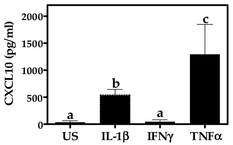
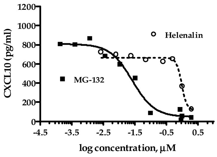
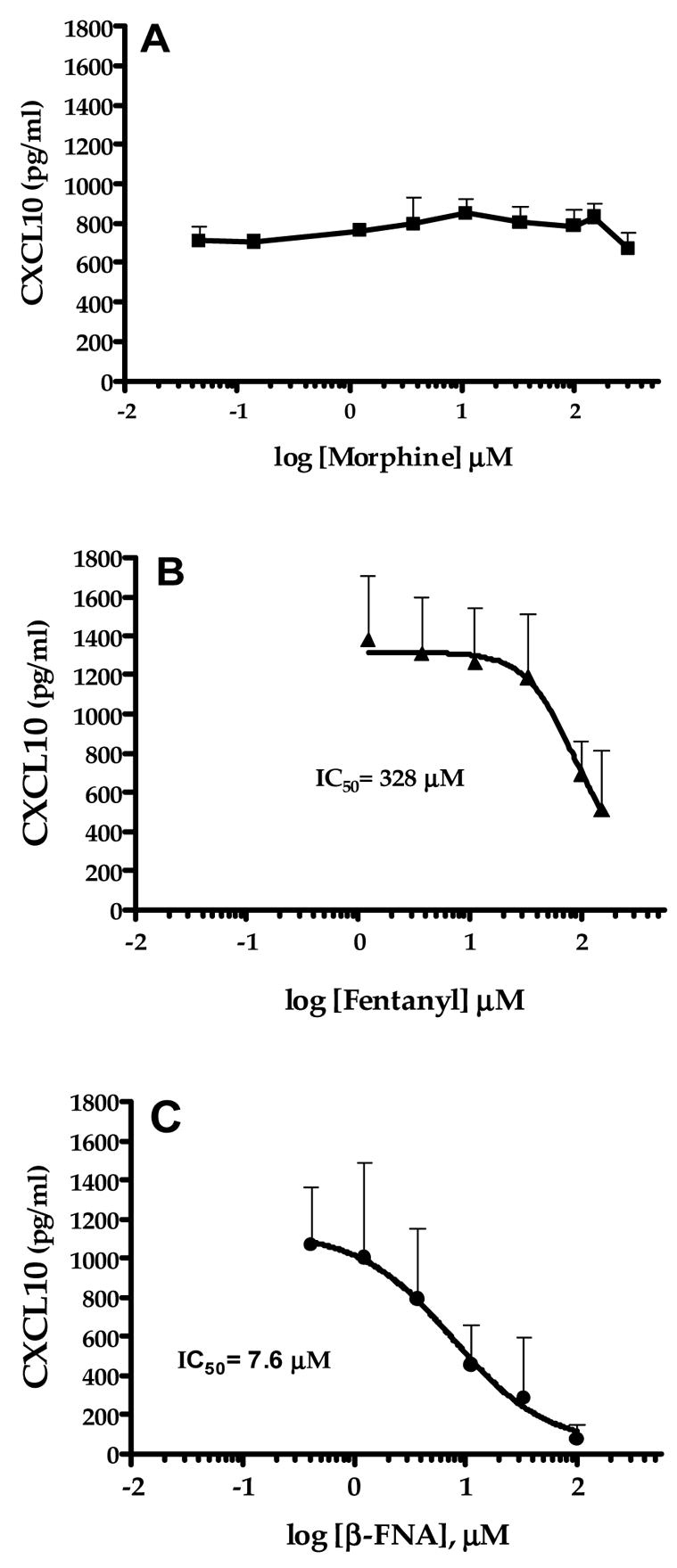
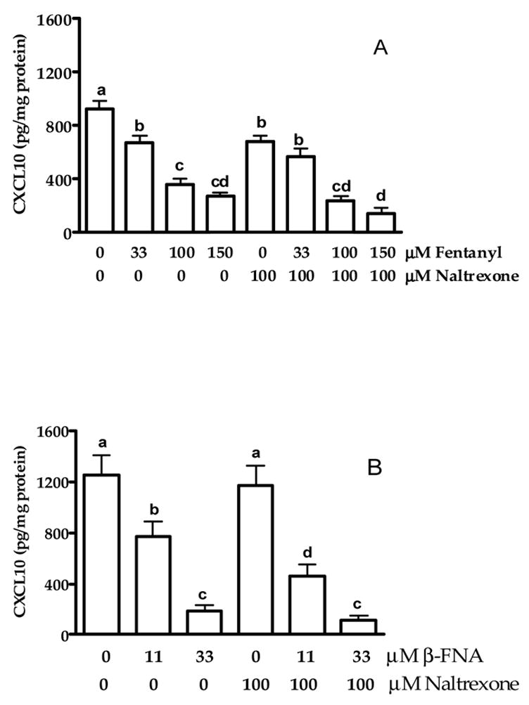
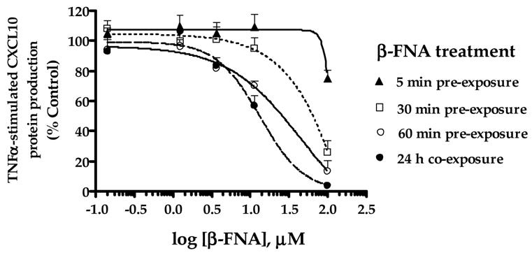
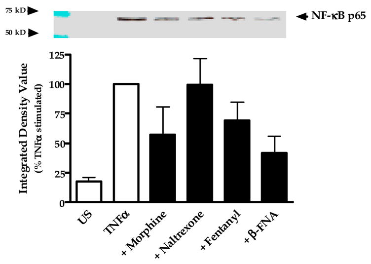
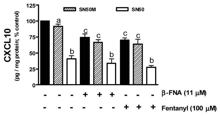
Emerging evidence indicates that neuroinflammatory responses in astroglia, including chemokine expression, are altered by opioids. Astroglial chemokines, such as CXCL10, are instrumental in response to many neuropathological insults. Opioid mediated disruption of astroglial CXCL10 expression may be detrimental in opioid abusers or patients receiving acute opioid therapy. We have characterized the in vitro effects of opioids on CXCL10 protein expression in human astroglial (A172) cells. The proinflammatory cytokine, tumor necrosis factor (TNF)alpha induced CXCL10 expression in A172 cells. Using MG-132, helenalin and SN50 [inhibitors of the transcription factor, nuclear factor (NF)-kappaB], we determined that NF-kappaB activation is instrumental in TNFalpha-induced CXCL10 expression in A172 astroglia. Morphine exposure during the 24 h TNFalpha stimulation period did not alter CXCL10 expression. However, fentanyl, a more potent mu-opioid receptor (MOR) agonist, inhibited TNFalpha-induced CXCL10 expression. Interestingly, neither the non-selective opioid receptor antagonist, naltrexone nor beta-funaltrexamine (beta-FNA), a highly selective MOR antagonist, blocked fentanyl mediated inhibition of TNFalpha-induced CXCL10 expression. Rather, beta-FNA dose-dependently inhibited TNFalpha-induced CXCL10 expression with a greater potency than that observed for fentanyl. Immunoblot analysis indicated that morphine, fentanyl and beta-FNA each reduced TNFalpha-induced nuclear translocation of NF-kappaB p65. These data show that beta-FNA and fentanyl inhibit TNFalpha-induced CXCL10 expression via a MOR-independent mechanism. Data also suggest that inhibition of TNFalpha-induced CXCL10 expression by fentanyl and beta-FNA is not directly related to a reduction in NF-kappaB p65 nuclear translocation. Further investigation is necessary in order to fully elucidate the mechanism through which these two opioid compounds inhibit CXCL10 expression. Understanding the mechanism by which chemokine expression is suppressed, particularly by the opioid antagonist, beta-FNA, may provide insights into the development of safe and effective treatments for neuroinflammation.


Figures

Fig. 1
Fig. 1
Cytokine-induced CXCL10 protein expression in A172 cells. Astroglial cells were unstimulated (US) or exposed for 24 h to human recombinant TNFα, IL-1β, or IFN-γ (5 ng/ml). A standard dual-antibody solid phase immunoassay (ELISA) was used for quantitation of secreted CXCL10 in cell culture supernatants. Data represent mean + S.E.M. of duplicate measures from 3 independent experiments. Data points with any common letters above them are not statistically different from each other as determined by one-way analysis of variance (ANOVA) with Neuman-Kuels’ multiple comparisons.
Fig. 2
Fig. 2
Inhibitors of NF-κB activation reduce TNFα-stimulated CXCL10 protein expression in A172 cells. Astroglial cells were exposed for 24 h to human recombinant TNFα (5 ng/ml) in the presence or absence of MG-132 (0.14 nM-2.0 μM; carbobenzoxy-L-leucyl-L-leucyl-L-leucinal) or the sesquiterpene lactone, helenalin (2.7 nM-2.0 μM). A standard dual-antibody solid phase immunoassay (ELISA) was used for quantitation of secreted CXCL10 in cell culture supernatants. Data points represent the mean + S.E.M. of duplicate measures from 2–7 independent experiments. Curves are from nonlinear regression in which the Hill slope was not constrained. The IC50 value and 95% confidence interval (CI) for MG-132 are 26 nM (95% CI: 17–41 nM) and 969 nM (95% CI: 754 nM-1.25 μM) for helenalin.
Fig. 3
Fig. 3
Modulation of TNFα-stimulated CXCL10 protein expression in A172 cells by selected opioids. Astroglial cells were exposed for 24 h to human recombinant TNFα (5 ng/ml) in the presence or absence of morphine (0.05–150 μM), fentanyl (1.2–150 μM) or β-funaltrexamine (β-FNA; 0.14–100 μM). A standard dual-antibody solid phase immunoassay (ELISA) was used for quantitation of secreted CXCL10 in cell culture supernatants. Data points represent the mean + S.E.M. of duplicate measures from 2–5 independent experiments. Curves are from nonlinear regression analysis in which the Hill slope was not constrained. The IC50 values are presented for each curve and the 95% confidence interval (CI) for each opioid are: morphine = not determined, fentanyl = 14.5–522.1 μM and β-FNA = 2.1–27.5 μM.
Fig. 4
Fig. 4
Effects of opioids on viability of TNFα stimulated astroglial cells. A172 cells were exposed for 24 h to human recombinant TNFα (5 ng/ml) in the presence or absence of morphine (1.23–100 μM), fentanyl (1.23–100 μM), naltrexone (1.23–100 μM) or β-funaltrexamine (β-FNA; 1.23–40 μM). Cell viability was determined by the MTT assay. Data represent mean + S.E.M. of duplicate or triplicate measures from 3 independent experiments. One-way analysis of variance (ANOVA) with Neuman-Kuels’ multiple comparisons indicated no significant opioid effects.
Fig. 5
Fig. 5
Modulation of (A) fentanyl- and (B) β-FNA-mediated inhibition of TNFα-stimulated CXCL10 protein expression in A172 cells by naltrexone. Astroglial cells were exposed for 24 h to human recombinant TNFα (5 ng/ml) in the presence or absence of fentanyl (33, 100 and 150 μM), β-funaltrexamine (β-FNA; 11 and 33 μM) and naltrexone (100 μM). A standard dual-antibody solid phase immunoassay (ELISA) was used for quantitation of secreted CXCL10 in cell culture supernatants. Data points represent the mean + S.E.M. of triplicate measures from 2 independent experiments (panel A) and quadruplicate measures from 3 independent experiments (panel B). Data points with any common letters above them are not statistically different from each other as determined by one-way analysis of variance (ANOVA) with Neuman-Kuels’ multiple comparisons.
Fig. 6
Fig. 6
Time-course effects of β-FNA on TNFα-stimulated CXCL10 protein expression in A172 cells. Astroglial cells were either co-exposed to β-funaltrexamine (β-FNA; 0.14–100 μM) and human recombinant TNFα (5 ng/ml) for 24 h (●) or pre-exposed to β-FNA for 5 (▲), 30 (□) or 60 (○) min, followed by washout of the drug, and subsequent exposure to TNFα for 24 h. A standard dual-antibody solid phase immunoassay (ELISA) was used for quantitation of secreted CXCL10 in cell culture supernatants. Data for each experiment were transformed to percent control (TNFα stimulation in the absence of β-FNA). Data points represent the mean + S.E.M. of duplicate measures from 3 independent experiments. Curves are resultant from nonlinear regression analysis in which the Hill slope was not constrained.
Fig. 7
Fig. 7
Modulation of NF-κB p65 nuclear translocation by opioids. Astroglial cells were exposed for 0.5 h to human recombinant TNFα (5 ng/ml) in the presence or absence of 33 μM morphine, naltrexone, fentanyl or β-funaltrexamine. Nuclear fractions were collected and NF-κB p65 protein levels assessed by immunoblot analysis of 10 μg total protein. β-tubulin levels were measured and used as loading controls. Images of the blots were digitally captured, and then integrated density values (IDV) were obtained using ImageJ software (NIH). β-tubulin band densities were used to normalize the densities of the NF-κB bands. Data points represent the mean + S.E.M. of the IDV obtained from 4 independent experiments (normalized for each experiment as percent stimulated control); the blot is representative of the 4 independent experiments.
Fig. 8
Fig. 8
Inhibition of TNFα stimulated CXCL10 expression by the NF-κB inhibitor SN50 and selected opioids β-FNA and fentanyl. Astroglial cells were preincubated for 1 h with SN50 (50 μM) or the mutant peptide SN50M (50 μM). Cells were then stimulated with TNFα (5 ng/ml) in the presence or absence of β-FNA (11 μM) or fentanyl (100 μM) for 24 h. A standard dual-antibody solid phase immunoassay (ELISA) was used for quantitation of secreted CXCL10 in cell culture supernatants. Data points represent the mean + S.E.M. of duplicate measures from 3 independent experiments. Data points with any common letters above them are not statistically different from each other as determined by one-way analysis of variance (ANOVA) with Neuman-Kuels’ multiple comparisons.

Similar articles

Cited by

References

    1. Bajetto A, Bonavia R, Barbero S, Schettini G. J Neurochem. 2002;82:1311–1329. - PubMed
    1. Baumhaker Y, Gafni M, Keren O, Sarne Y. Mol Pharmacol. 1993;44:461–467. - PubMed
    1. Beech JS, Reckless J, Mosedale DE, Grainger DJ, Williams SC, Menon DK. J Cereb Blood Flow Metab. 2001;21:683–689. - PubMed
    1. Belperio JA, Keane MP, Arenberg DA, Addison CL, Ehlert JE, Burdick MD, Strieter RM. J Leukoc Biol. 2000;68:1–8. - PubMed
    1. Borner C, Kraus J, Schroder H, Ammer H, Hollt V. Mol Pharmacol. 2004;66:1719–1726. - PubMed

Publication types

MeSH terms