. 2007 May 15;17(10):834-43.
doi: 10.1016/j.cub.2007.04.036. Epub 2007 May 3.

Overexpressing centriole-replication proteins in vivo induces centriole overduplication and de novo formation

Affiliations

Overexpressing centriole-replication proteins in vivo induces centriole overduplication and de novo formation

Nina Peel et al. Curr Biol. .

Abstract

Background: Centrosomes have important roles in many aspects of cell organization, and aberrations in their number and function are associated with various diseases, including cancer. Centrosomes consist of a pair of centrioles surrounded by a pericentriolar matrix (PCM), and their replication is tightly regulated. Here, we investigate the effects of overexpressing the three proteins known to be required for centriole replication in Drosophila-DSas-6, DSas-4, and Sak.

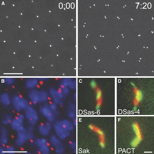
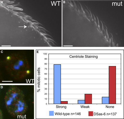
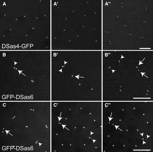
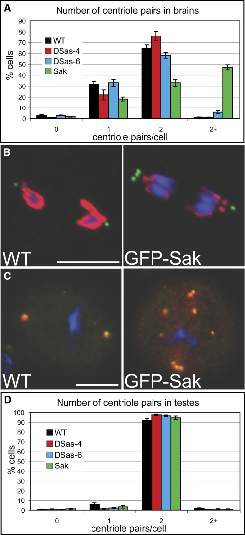
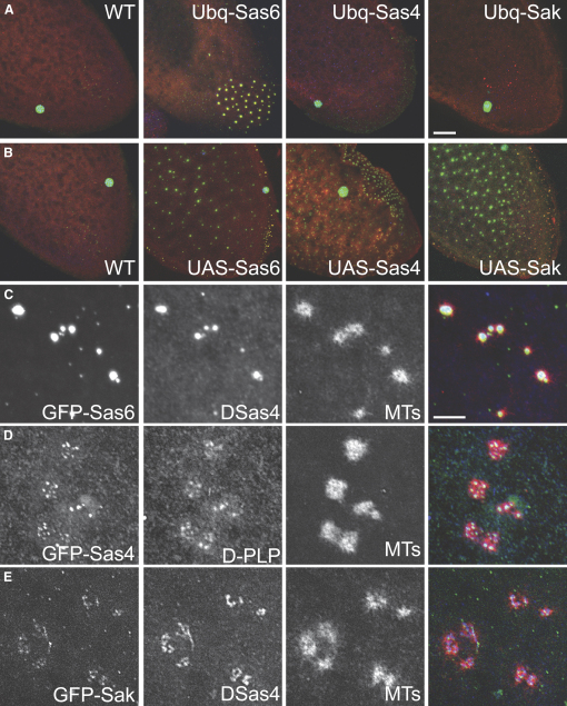
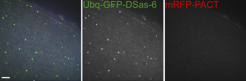
Results: By directly observing centriole replication in living Drosophila embryos, we show that the overexpression of GFP-DSas-6 can drive extra rounds of centriole replication within a single cell cycle. Extra centriole-like structures also accumulate in brain cells that overexpress either GFP-DSas-6 or GFP-Sak, but not DSas-4-GFP. No extra centrioles accumulate in spermatocytes that overexpress any of these three proteins. Most remarkably, the overexpression of any one of these three proteins results in the rapid de novo formation of many hundreds of centriole-like structures in unfertilized eggs, which normally do not contain centrioles.

Conclusions: Our data suggest that the levels of centriolar DSas-6 determine the number of daughter centrioles formed during centriole replication. Overexpression of either DSas-6 or Sak can induce the formation of extra centrioles in some tissues but not others, suggesting that centriole replication is regulated differently in different tissues. The finding that the overexpression of DSas-4, DSas-6, or Sak can rapidly induce the de novo formation of centriole-like structures in Drosophila eggs suggests that this process results from the stabilization of centriole-precursors that are normally present in the egg.


Figures

Figure 1
Figure 1
DSas-6 Is a Centriolar Protein (A) GFP-DSas-6 associates with centrioles in living Drosophila embryos. Time in min:s is shown in each panel. In interphase (0:00), two centriole pairs are associated with each nucleus, but these pairs can only be resolved as single dots at this stage. These centriole pairs separate from one another in telophase (7:20). (B) The localization of GFP-DSas-6 (pseudocolored red) in whole-mount third instar larval brains. DNA is shown in blue. The centrioles in these cells do not associate with any PCM in interphase , so the presence of one or two GFP-DSas-6 dots in every cell indicates that the protein is centriolar, rather than centrosomal. (C–F) The localization of GFP-DSas-6 (C), DSas-4-GFP (D), GFP-Sak (E), and GFP-PACT (F) (green) in spermatocyte centrioles. The centrioles are stained with GTU88 (red). Note how GTU88 and GFP-PACT evenly label the entire centriole, whereas the other GFP-fusions are concentrated at the proximal and distal ends. Scale bars represent 10 μm in (A), 5 μm in (B), and 2 μm (C)–(F).
Figure 2
Figure 2
DSas-6 Is Required for Centriole Replication (A and B) The distribution of basal bodies (revealed here with GFP-PACT) in WT (A) and mutant (B) third antennal segments. Basal bodies are present at the base of each sensory bristle in WT antenna (arrow) but are undetectable in mutant antenna. (C and D) The localization of the centriolar marker DSas-4 (red) and the PCM marker Cnn (green) in WT (C) and DSas-6 mutant (D) mitotic larval brain cells. No centrioles or centrosomes are detectable in the mutant cells. (E) Quantitation of centriole numbers (D-PLP-positive dots) in WT and DSas-6 mutant mitotic cells (see also Figure S3). More than 70% of mutant cells have no detectable centrioles. Scale bars represent 10 μm.
Figure 3
Figure 3
Centriole Overduplication in Embryos Expressing GFP-DSas-6 (A) In embryos expressing DSas-4-GFP, centrioles replicate normally. (B) In the GFP-DSas-6-expressing embryo shown here, the two centrioles marked with an arrow and arrowhead initially separate from one another at telophase (B). During the next interphase, one of the centrioles produces an extra centriole (arrowheads, [B′]). At the next telophase, both of these “extra” centrioles replicate normally and separate in synchrony with the other centrioles in the embryo (arrowheads, [B″]). (C) In the GFP-DSas-6 embryo shown here, two centrioles are highlighted (arrow and arrowhead) at telophase (C). In the next interphase, both of these centrioles produce an extra centriole (arrows and arrowheads, [C′]). The centriole at the bottom right (arrowheads) then replicates one more time to produce three centrioles (C″). See Movies S1 and S2. Scale bar represents 10 μm.
Figure 4
Figure 4
The Overexpression of GFP-DSas-6 and GFP-Sak Drives the Accumulation of Extra Centrioles in Third Instar Larval Brain Cells (A) Quantitation of centriole number (D-PLP-positive dots) in mitotic cells from third instar larval brains expressing different GFP-fusions. For each fusion protein n > 1200 cells (in total) from at least 4 different brain preparations. (B and C) WT or GFP-Sak-expressing mitotic brain cells were stained to reveal centrioles (D-PLP, green) and phospho-Histone H3 (red) (B) or centrioles (D-PLP, green) and γ-tubulin (red) (C). Note how the extra centrioles can cluster at the poles of the spindle (B) and can organize γ-tubulin (C), suggesting that they are at least partially functional. Scale bars represent 10 μm. (D) Quantitation of centriole number (GTU88-positive centrioles) in spermatocytes expressing different GFP-fusions (as indicated in the panel). For each fusion protein n > 750 cells (in total) from four different testes preparations. Error bars represent the standard error.
Figure 5
Figure 5
The High-Level Overexpression of Centriole-Replication Proteins Drives the De Novo Formation of Centriole-like Structures in Unfertilized Eggs (A) Unfertilized eggs laid by WT, Ubq-GFP-DSas-6, Ubq-DSas-4-GFP, and Ubq-GFP-Sak females showing the distribution of endogenous DSas-4 (red) and MTs (green). The expression of GFP-DSas-6 can induce the formation of a relatively small number of centriole-like structures. The MT structures in the other eggs are the MTs that surround the polar bodies. (B) Unfertilized eggs laid by WT, UAS-GFP-DSas-6, UAS-DSas-4-GFP, and UAS-Sak-GFP females. All of the eggs are filled with many centriole-like structures. (C and D) Higher-magnification views of the centriole-like structures formed in eggs laid by UAS-GFP-DSas-6 (C), UAS-DSas-4-GFP (D), and UAS-Sak-GFP (E) females. GFP fluorescence is shown in the left panels (green in merged image), endogenous DSas-4 or D-PLP in the middle panels (blue in merged image), and MTs in the right panels (red in merged image). Scale bars represent 25 μm in (A) and (B) and 5 μm in (C)–(E).
Figure 6
Figure 6
Centrioles Appear to be Inactivated Normally in Oocytes Expressing Centriole-Replication Proteins In stage 14 oocytes, some GFP-aggregates can still be detected in oocytes expressing any of these GFP-fusions (Ubq-DSas-6 is shown here), but none of these colocalize with mRFP-PACT, suggesting that they are not centrioles. Scale bar represents 10 μm.

Similar articles

Cited by

References

    1. Kellogg D.R., Moritz M., Alberts B.M. The centrosome and cellular organization. Annu. Rev. Biochem. 1994;63:639–674. - PubMed
    1. Nigg E.A. Centrosome aberrations: cause or consequence of cancer progression? Nat. Rev. Cancer. 2002;2:815–825. - PubMed
    1. Raff J.W. Centrosomes and cancer: lessons from a TACC. Trends Cell Biol. 2002;12:222–225. - PubMed
    1. Basto R., Lau J., Vinogradova T., Gardiol A., Woods C.G., Khodjakov A., Raff J.W. Flies without centrioles. Cell. 2006;125:1375–1386. - PubMed
    1. Uetake Y., Loncarek J., Nordberg J.J., English C.N., La Terra S., Khodjakov A., Sluder G. Cell cycle progression and de novo centriole assembly after centrosomal removal in untransformed human cells. J. Cell Biol. 2007;176:173–182. - PMC - PubMed

Publication types

MeSH terms

Substances

LinkOut - more resources