. 2007 Jul;81(14):7351-62.
doi: 10.1128/JVI.00554-07. Epub 2007 May 2.

Tumor necrosis factor activates a conserved innate antiviral response to hepatitis B virus that destabilizes nucleocapsids and reduces nuclear viral DNA

Affiliations

Tumor necrosis factor activates a conserved innate antiviral response to hepatitis B virus that destabilizes nucleocapsids and reduces nuclear viral DNA

Robyn Puro et al. J Virol. 2007 Jul.

Abstract

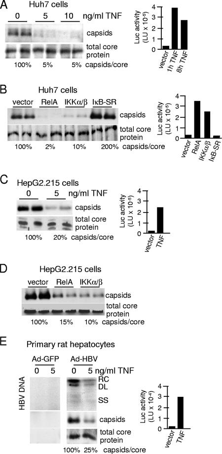
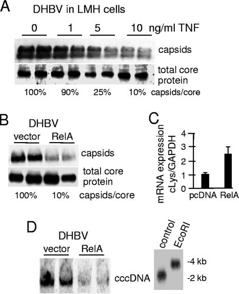
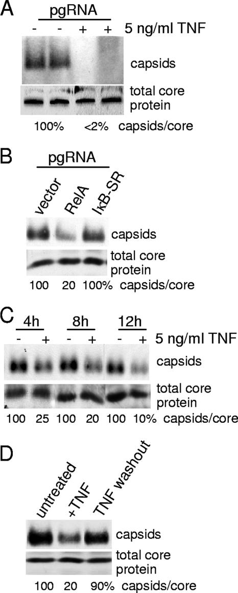
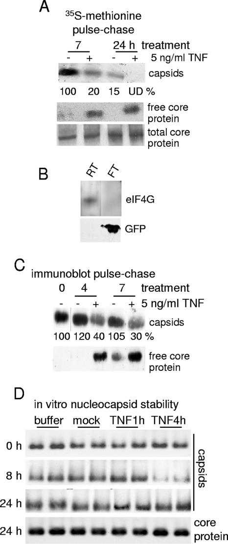
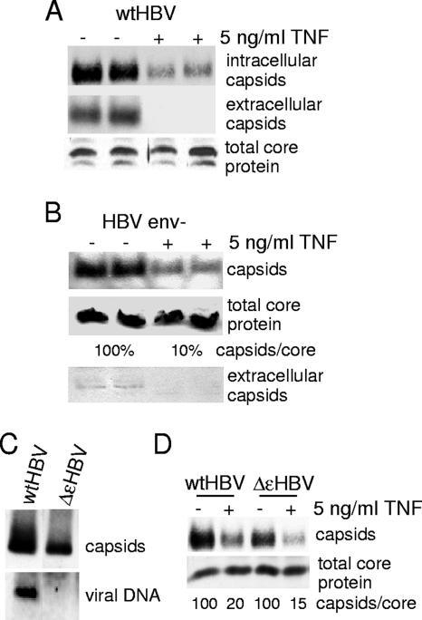
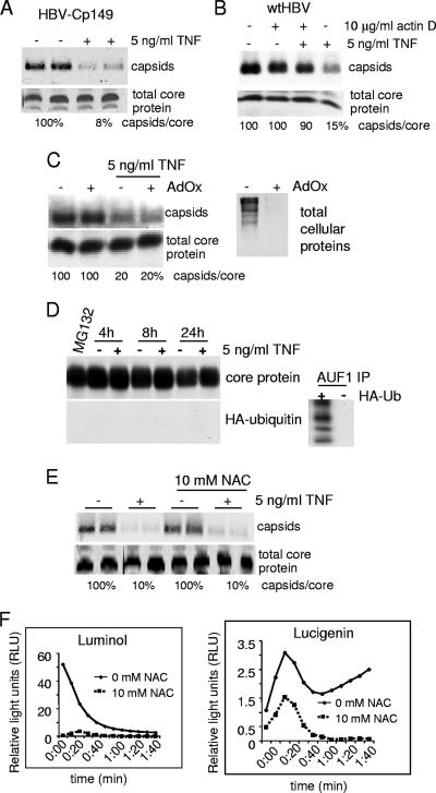
Tumor necrosis factor (TNF) is critical for the control of hepatitis B virus (HBV) in the clinical setting and in model systems. TNF induces noncytopathic suppression and clearance of HBV in animal models, possibly through reduction of viral nucleocapsids, but the mechanism is not well described. Here, we demonstrate the molecular mechanism and broad host range for TNF action against HBV. We show that TNF rapidly blocks HBV replication by promoting destabilization of preexisting cytoplasmic viral nucleocapsids containing viral RNA and DNA, as well as empty nucleocapsids. TNF destabilized human HBV nucleocapsids in a variety of human hepatocytic cell lines and in primary rat hepatocytes and also destabilized duck HBV (DHBV) nucleocapsids in chicken hepatocytic cells. Lysates from TNF-treated uninfected cells also destabilized HBV nucleocapsids in vitro. Moreover, inhibition of DHBV DNA replication by TNF blocks nuclear accumulation of the viral transcription template, maintenance of which is essential for the establishment and maintenance of chronic infection. We show that TNF destabilization of HBV nucleocapsids does not involve ubiquitination or methylation of the viral core protein and is not mediated by the nitric oxide free radical arm of the TNF pathway. These results define a novel antiviral mechanism mediated by TNF against multiple types of HBVs in different species.


Figures

FIG. 1.
FIG. 1.
TNF downregulates human HBV nucleocapsid levels in different hepatocytic cells lines and primary hepatocytes. (A) Huh7 cells were transfected with an HBV genomic replicon plasmid and an NF-κB-dependent luciferase reporter and then treated 2 days later with 5 ng/ml TNF for up to 24 h. Cytoplasmic nucleocapsids were resolved by native agarose gel electrophoresis and subjected to immunoblot analysis with antibodies to HBcAg. Total core protein levels were quantified by SDS-PAGE and immunoblot analysis with antibodies to HBcAg. Luciferase activity was determined at the times shown using a luminometer, and the results of three experiments were averaged for presentation. (B) Huh7 cells were transfected with an HBV genomic replicon plasmid and vector alone or expression vectors for RelA, IKKα/β or IκB-SR, and an NF-κB-dependent luciferase reporter. Nucleocapsids and total core protein levels were assessed at 48 h as described above, in duplicate. Luciferase activities were determined at the same time and averaged from three independent experiments. (C) HepG2.215 cells were transfected with the NF-κB luciferase reporter and mock treated or treated with 5 ng/ml TNF for 24 h, and nucleocapsids/total core protein levels and luciferase activity were determined as described above. (D) HepG2.215 cells were transfected with vector alone or expression vectors for RelA or IKK-α/β, and nucleocapsids/total core protein levels were examined as described above. (E) Primary rat hepatocytes were purified, transfected with an NF-κB luciferase reporter plasmid, and transduced with an Ad vector at 50 particles per cell expressing a replicon of HBV (Ad-HBV) or control Ad-GFP for 3 days and then treated with 5 ng/ml human TNF for 3 days. Cytoplasmic viral nucleocapsids were purified from equal numbers of cells, and associated HBV DNA was detected by Southern blot analysis. Nucleocapsids and total core protein levels were assessed as described above. Luciferase activity was determined as described above. Results shown are representative of three independent experiments performed in duplicate. Abbreviations: RC, relaxed circular form; DL, double-strand linear form; SS, single-strand form; Luc, luciferase.
FIG. 2.
FIG. 2.
TNF and NF-κB downregulate levels of DHBV cytoplasmic nucleocapsids and nuclear cccDNA. (A) LMH cells (chicken hepatocytic cell line) were transfected with a DHBV replicon consisting of a trimer of head-to-tail genomic DNA and treated with 0, 1, 5, or 10 ng/ml TNF for 24 h. Cytoplasmic nucleocapsids were resolved by native agarose gel electrophoresis and subjected to immunoblot analysis with DHBcAg antibody. Total core protein levels were determined by SDS-PAGE followed by immunoblot analysis. All studies were performed in duplicate. (B) LMH cells were transfected with the DHBV replicon and either vector alone or a plasmid expressing RelA. Capsid and total core protein levels were determined as described above and values are presented as the means of three studies. (C) mRNA was isolated from LMH cells cotransfected with the DHBV replicon and vector alone or RelA plasmid, and expression levels of chicken lysozyme were quantified and analyzed by quantitative real-time PCR. (D) LMH cells were cotransfected with the DHBV replicon and vector alone or with RelA plasmid. cccDNA was isolated from equal numbers of cell nuclei, and levels were analyzed by Southern hybridization analysis as described in Materials and Methods. The authenticity of cccDNA was established by restriction enzyme digestion and an electrophoretic mobility shift change from 2 kb (cccDNA) to 4 kb (linear DNA) with EcoRI, which linearizes the cccDNA. Results shown are representative of at least three independent experiments performed in duplicate.
FIG. 3.
FIG. 3.
TNF acts posttranscriptionally and rapidly to downregulate HBV nucleocapsid levels. (A) HepG2 cells were transfected with in vitro-transcribed pgRNA (see Materials and Methods for details). Following transfection, cells were treated with 5 ng/ml TNF for 24 h in duplicate studies as shown or (B) secondarily transfected with a vector control or expression plasmids for RelA or IκB-SR. Nucleocapsids were isolated and analyzed by native agarose gel electrophoresis and immunoblotting, and total core protein was analyzed by SDS-PAGE and immunoblotting using an anti-HBcAg antibody. (C) HepG2 cells were transfected with an HBV replicon plasmid for 2 days and then treated with 5 ng/ml TNF for 4, 8, or 12 h. Cytoplasmic nucleocapsids and total core protein levels were determined as described above. (D) HepG2 cells were transfected with an HBV replicon as described above, treated with 5 ng/ml of TNF for 24 h, washed, and cultured for another 24 h. Total core protein and nucleocapsid levels were analyzed by electrophoresis and immunoblotting. Results shown are representative of at least three independent experiments.
FIG. 4.
FIG. 4.
TNF mediates disassembly of preexisting nucleocapsids. (A) HepG2.215 cells were subjected to pulse-chase analysis with [35S]methionine-cysteine (see Materials and Methods for details). Nucleocapsids were separated from “free” core protein (nonnucleocapsid protein) using Centricon-100 filtration with a >100-kDa molecular size exclusion limit, and nucleocapsids, free core proteins, and total core protein in lysates were immunoprecipitated using an anti-HBcAg antibody. Capsid data were quantified by densitometry and expressed as normalized to the untreated 7-h mock control lane. UD, undetectable. (B) Centricon-100 analysis control data are shown, demonstrating the retention of eukaryotic initiation factor 4 subunit G (eIF4G), a 220-kDa protein, in the retentate fraction (RT) and the passage of 25-kDa green fluorescent protein (GFP) in the flowthrough fraction (FT). (C) HepG2.215 cells were unlabeled and analyzed as described above. Nucleocapsids and free core protein levels were determined by Centricon-100 filtration as described above, and proteins were detected by immunoblot analysis. Capsid data were quantified as described above and normalized to the untreated (0-h) capsid level. (D) HepG2 cells transfected with HBV replicons and untransfected HepG2 cells were left untreated or were treated with 5 ng/ml TNF for 1 h or 4 h. Cells were harvested and lysed by Dounce homogenization, and crude extracts from HBV-transfected cells as a source of viral nucleocapsids were incubated at 30°C with lysates obtained from the untransfected HepG2 cells. Nucleocapsid integrity was assessed over a 24-h period by native agarose gel electrophoresis and immunoblot analysis, in duplicate samples. Results shown are representative of at least three independent experiments.
FIG. 5.
FIG. 5.
TNF promotes destabilization and not premature secretion of mature, empty, and nonenveloped nucleocapsids. (A) HepG2 cells were transfected with a wild-type HBV replicon (wtHBV) plasmid and then treated with 5 ng/ml TNF for 24 h. Extracellular and intracellular nucleocapsids were collected and analyzed by native agarose gel electrophoresis and immunoblot analysis in duplicate. Total core protein levels were determined by SDS-PAGE and immunoblot analysis. (B) HepG2 cells were transfected with an env HBV replicon (HBV env-) that cannot produce HBsAgs or secrete mature enveloped nucleocapsids and then treated with 5 ng/ml TNF for 24 h. Cytoplasmic and extracellular nucleocapsids and total core protein levels were analyzed as described above. (C) Cytoplasmic nucleocapsids were purified from HepG2 cells transfected with wild-type HBV or ΔɛHBV replicons and nucleocapsid and encapsidated viral DNA were examined by immunoblotting and Southern blot DNA analysis. (D) HepG2 cells were transfected with either a wild-type HBV replicon or ΔɛHBV, a replicon mutated in the epsilon region that cannot encapsidate pgRNA into nucleocapsids. Cells were treated with 5 ng/ml TNF, and nucleocapsids and total core protein were analyzed as described above. Results shown are representative of at least three independent experiments.
FIG. 6.
FIG. 6.
TNF disruption of nucleocapsids does not require the regulatory C-terminal region of core protein but does require cellular transcription. (A) HepG2 cells were transfected with a Cp149 HBV replicon (HBV-Cp149) that lacks the C terminus of core protein from amino acid 149 and then treated with 5 ng/ml TNF for 24 h. Nucleocapsids and total core protein were analyzed as described earlier. wtHBV, wild-type HBV replicon. (B) HepG2 cells were transfected with a wild-type HBV replicon for 2 days and pretreated with actinomycin D (actin D) for 30 min, followed by the addition of 5 ng/ml TNF for 4 h. Nucleocapsids and total core protein were analyzed as described earlier. Results shown are representative of at least three independent experiments. (C) HepG2 cells were transfected with the HBV replicon plasmid, and then 24 h later cells were pretreated with 100 μM for 24 h followed by the addition of 5 ng/ml TNF for 6 h. Nucleocapsids and total core protein were analyzed as described above. The inhibition of protein methylation by AdOx was examined by labeling cells with l-[methyl-3H]-methionine in the presence or absence of AdOx followed by SDS-PAGE and fluorography. (D) HepG2 cells were cotransfected with the HBV replicon for 48 h and with an HA-ubiquitin expression plasmid and then treated with 5 ng/ml TNF for 4, 8, or 24 h or incubated with 10 μM of proteasome inhibitor MG132, and AUF1 or HA was immunoprecipitated and analyzed by SDS-PAGE and immunoblot analysis with anti-HA or anti-HBcAg antibodies. (E) HepG2 cells were transfected with HBV replicon DNA, treated 24 h later with 10 mM NAC for 2 h, and then incubated with 5 ng/ml TNF for 6 h (in the presence of NAC). Nucleocapsid and total core protein levels were assayed in duplicate as described above. (F) HepG2 cells were transfected with HBV replicons, and then at 24 h cells were left untreated or were treated with 10 mM NAC to block ROS and incubated with luminol or lucigenin. Cells were then exposed to 0.1% H2O2, and free radical production was measured by luminol or lucigenin fluorescence. Data are expressed in relative light units.

Similar articles

Cited by

References

    1. al-Wabel, A., M. al-Janadi, and S. Raziuddin. 1993. Cytokine profile of viral and autoimmune chronic active hepatitis. J. Allergy Clin. Immunol. 92:902-908. - PubMed
    1. Ando, K., L. G. Guidotti, S. Wirth, T. Ishikawa, G. Missale, T. Moriyama, R. D. Schreiber, H. J. Schlicht, S. N. Huang, and F. V. Chisari. 1994. Class I-restricted cytotoxic T lymphocytes are directly cytopathic for their target cells in vivo. J. Immunol. 152:3245-3253. - PubMed
    1. Baron, J. L., L. Gardiner, S. Nishimura, K. Shinkai, R. Locksley, and D. Ganem. 2002. Activation of a nonclassical NKT cell subset in a transgenic mouse model of hepatitis B virus infection. Immunity 16:583-594. - PubMed
    1. Barrasa, M. I., J. T. Guo, J. Saputelli, W. S. Mason, and C. Seeger. 2001. Does a cdc2 kinase-like recognition motif on the core protein of hepadnaviruses regulate assembly and disintegration of capsids? J. Virol. 75:2024-2028. - PMC - PubMed
    1. Biermer, M., R. Puro, and R. J. Schneider. 2003. Tumor necrosis factor alpha inhibition of hepatitis B virus replication involves disruption of capsid integrity through activation of NF-κB. J. Virol. 77:4033-4042. - PMC - PubMed

Publication types

MeSH terms

LinkOut - more resources