. 2007 Jul;18(7):2511-24.
doi: 10.1091/mbc.e06-11-0995. Epub 2007 May 2.

The transmembrane domain of acid trehalase mediates ubiquitin-independent multivesicular body pathway sorting

Affiliations

The transmembrane domain of acid trehalase mediates ubiquitin-independent multivesicular body pathway sorting

Ju Huang et al. Mol Biol Cell. 2007 Jul.

Abstract

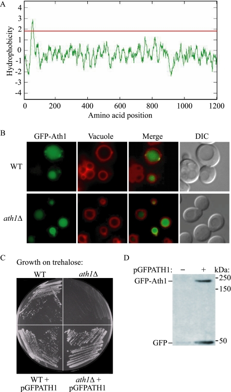
Trehalose serves as a storage source of carbon and plays important roles under various stress conditions. For example, in many organisms trehalose has a critical function in preserving membrane structure and fluidity during dehydration/rehydration. In the yeast Saccharomyces cerevisiae, trehalose accumulates in the cell when the nutrient supply is limited but is rapidly degraded when the supply of nutrients is renewed. Hydrolysis of trehalose in yeast depends on neutral trehalase and acid trehalase (Ath1). Ath1 resides and functions in the vacuole; however, it appears to catalyze the hydrolysis of extracellular trehalose. Little is known about the transport route of Ath1 to the vacuole or how it encounters its substrate. Here, through the use of various trafficking mutants we showed that this hydrolase reaches its final destination through the multivesicular body (MVB) pathway. In contrast to the vast majority of proteins sorted into this pathway, Ath1 does not require ubiquitination for proper localization. Mutagenesis analyses aimed at identifying the unknown targeting signal revealed that the transmembrane domain of Ath1 contains the information sufficient for its selective sequestration into MVB internal vesicles.


Figures

Figure 1.
Figure 1.
Ath1 is localized in the vacuolar lumen in S. cerevisiae. (A) A hydropathy plot was created by the Kyte-Doolittle algorithm (Kyte and Doolittle, 1982) based on the amino acid sequence of Ath1, and one transmembrane domain was predicted at amino acids 51–69. Peaks with scores greater than 1.8 (line) indicate a possible transmembrane region. (B) Wild-type (WT; BY4742) and ath1Δ strains were transformed with a plasmid (pGFPATH1) encoding a GFP-Ath1 fusion protein controlled by a constitutively active, strong TPI1 promoter. Yeast cells were grown in SMD-URA medium to log phase, treated with FM 4-64 to stain the vacuole and observed with a fluorescence microscope as described in Materials and Methods. DIC, differential interference contrast. (C) WT, ath1Δ, or the same strains harboring the pGFPATH1 plasmid were streaked on SMT-URA medium in which trehalose is the sole carbon source. The plate was grown at 30°C for 7 d. (D) The ath1Δ strain harboring either the pRS416 empty vector (−) or pGFPATH1 (+) plasmid were collected at log phase and whole cell extracts were subjected to SDS-PAGE. GFP or GFP-Ath1 were detected by immunoblot probed with monoclonal anti-GFP antibody. Molecular mass (kDa) of protein standards is indicated on the right side of the blot.
Figure 2.
Figure 2.
Acid trehalase activity is primarily localized within the vacuole. (A) Vacuoles from the same amount of cells of the wild-type (WT; BY4742) and ath1Δ strains were prepared and assayed as described in Materials and Methods. Acid trehalase activity from the total cell lysate (Total) as well as the vacuole fraction (Vacuole) was measured. Final vacuolar Ath1 activity was calculated as the activity present in the vacuole fraction divided by the level of vacuole recovery according to the percentage of α-mannosidase in the vacuole fraction shown in B. The wild-type total Ath1 activity was set at 100%, and the other values were normalized accordingly. (B) Enzyme assays of α-mannosidase, α-glucosidase, and NADPH cytochrome c reductase were carried out as described in Materials and Methods. The percentages of enzyme activity in the vacuole fraction compared with the total loaded cell lysate from both wild-type and ath1Δ strains were plotted. All experiments were repeated three times independently. Error bar, SD among the repeats.
Figure 3.
Figure 3.
Ath1 is delivered to the vacuole through the MVB pathway. GFP-Ath1 was either expressed under the TPI1 promoter (A) or under its endogenous promoter (B) in wild-type (WT), atg1Δ, apm3Δ, vps4Δ, vps27Δ, and end3Δ strains. Cells overexpressing GFP-Ath1 were grown in SMD-URA medium to log phase, whereas cells expressing the endogenous level of GFP-Ath1 were grown to stationary phase to induce Ath1 expression. Vacuoles were labeled with FM 4-64, and GFP-Ath1 localization was observed with a fluorescence microscope.
Figure 4.
Figure 4.
Ath1 is a glycosylated, type II transmembrane protein. (A) A whole cell extract from wild-type cells harboring the pGFPAth1 plasmid was prepared and treated without (−) or with (+) endoglycosidase H as described in Materials and Methods. Samples were resolved by SDS-PAGE and subjected to Western blot probed with anti-GFP, anti-Prc1, and anti-Ape1 antibody or antisera (faster migrating species corresponding to free GFP were not detected). Protein bands of precursor Ape1 (prApe1) and mature Ape1 (mApe1) are indicated. (B) Schematic diagram of the localization and cleavage patterns of the GFP-Ath1 chimera in wild-type (WT), vps4Δ, and pep4Δ vps4Δ strains. The predicted molecular mass of the GFP-containing species is indicated. (C) Cells from wild-type (WT), vps4Δ, and pep4Δ vps4Δ strains were transformed with the pRS416 empty vector (−) or the pGFPAth1 plasmid (+). Whole cell extracts were analyzed by immunoblotting with anti-GFP antibody. The asterisks indicate nonspecific bands.
Figure 5.
Figure 5.
Ath1 is a transmembrane protein with its C terminus in the lumen. (A) Wild-type (WT) and Ath1-HA (YJH1) strains were grown to stationary phase, and the cells were converted to spheroplasts. An aliquot was removed for the total sample, and the remainder was fractionated into supernatant (S13) and pellet (P13) fractions according to Materials and Methods. The Ath1-HA fusion protein was mostly recovered in the P13 fraction. The cytosolic protein Pgk1 serves as a control to monitor spheroplast lysis and separation of the fractions. (B) The Ath1-HA pep4Δ vps4Δ strain (YJH35) was fractionated as above and the P13 fraction was resuspended in either high-salt buffer (1 M KCl), high-pH buffer (0.1 M Na2CO3, pH 11.0), or detergent (1% Triton X-100). After treatment, samples were centrifuged at 13,000 × g for 5 min and separated into supernatant (S) and pellet (P) fractions. Antibodies against HA and Pho8 were used to detect the Ath1-HA fusion protein and Pho8. (C) The P13 fraction from B was resuspended in PS200 buffer in the presence (+) or absence (−) of proteinase K and Triton X-100, as indicated, then resolved by SDS-PAGE, and subjected to Western blot with anti-HA antibody.
Figure 6.
Figure 6.
Ath1 is sorted into the MVB pathway in an ubiquitin-independent manner. (A) The ath1Δ strain was transformed with pGFPATH1 plasmids encoding Ath1 with the indicated mutations of lysine to arginine in the cytosolic domain. The localization of the chimeric proteins was monitored by fluorescence microscopy. (B) The pGFPATH1, pGFPPHM5, or pGFPSNA3 plasmids were transformed into tul1Δ, bsd2Δ (XGY5), bsd2Δ tul1Δ (YJH38), and fab1Δ mutant strains. Intracellular localization of all GFP-fused proteins was observed by fluorescence microscopy. (C) bsd2Δ cells expressing GFP-Ath1 were grown in SMD-URA media. Cells were collected at early growth phase (OD600 = 0.8–1) and late growth phase (OD600 = 2–3) and subjected to fluorescence microscopy. Differential interference contrast (DIC) images are presented to the right of the GFP images.
Figure 7.
Figure 7.
Ath1 is ubiquitinated on its cytosolic domain. (A) Wild-type (WT) and Ath1-HA (YJH1) cells were immunoprecipitated with anti-HA antibody as described in Materials and Methods. The immunoprecipitate was subjected to SDS-PAGE and immunoblotted with anti-HA or anti-Ub antibody. The precipitated Ath1-HA protein was detected with anti-Ub antibody, indicated as Ub-Ath1-HA. (B) doa4Δ (MHY623) cells were transformed with plasmids expressing HA-Ub alone, GFP-Ath1 alone or together with HA-Ub, or GFPAth1K2,27,37R (GFP-Ath1KR) alone or together with HA-Ub. Cell lysates were immunoprecipitated with anti-HA antibody and then immunoblotted with anti-GFP and anti-Cps1 antibody or antiserum. The asterisk indicates a nonspecific band.
Figure 8.
Figure 8.
The transmembrane domain of Ath1 contains a sufficient signal for sorting into the MVB pathway. (A) The pGFPATH1ΔC, pGFPATH1ΔN, and pGFPATH1TM constructs (shown schematically on the right) were transformed into ath1Δ cells separately to express GFP-tagged Ath1 containing the N terminus and transmembrane domains, the C terminus and transmembrane domains, or only the transmembrane domain, respectively. All of the altered proteins were still localized within the vacuole lumen. (B) The fab1Δ, tul1Δ, bsd2Δ tul1Δ, and wild-type (WT) strains expressing either wild-type GFP-Phm5 or GFP-Phm5/Ath1TM, in which the original transmembrane domain of Phm5 was replaced with the transmembrane domain of Ath1, were examined by fluorescence microscopy. Differential interference contrast (DIC) images are presented to the right of the GFP images in A and B. (C) Wild-type cells transformed with plasmids expressing GFP-Ath1ΔN, GFP-Ath1polarmut (in which four polar amino acid residues in the transmembrane domain had been replaced with hydrophobic residues), or GFP-Ath1ΔNpolarmut mutant proteins were monitored by fluorescence microscopy. Images from 500 randomly selected cells containing a GFP signal from each strain were analyzed, and cells that showed a weak or partial vacuolar membrane fluorescent signal were counted. The percentage of cells displaying vacuolar membrane fluorescence for each construct is shown beneath the corresponding image. The white arrowhead points to the ER.

Similar articles

Cited by

References

    1. Alizadeh P., Klionsky D. J. Purification and biochemical characterization of the ATH1 gene product, vacuolar acid trehalase, from Saccharomyces cerevisiae. FEBS Lett. 1996;391:273–278. - PubMed
    1. Avaro S., Belgareh-Touze N., Sibella-Arguelles C., Volland C., Haguenauer-Tsapis R. Mutants defective in secretory/vacuolar pathways in the EUROFAN collection of yeast disruptants. Yeast. 2002;19:351–371. - PubMed
    1. Babst M. A protein's final ESCRT. Traffic. 2005;6:2–9. - PubMed
    1. Babst M., Sato T. K., Banta L. M., Emr S. D. Endosomal transport function in yeast requires a novel AAA-type ATPase, Vps4p. EMBO J. 1997;16:1820–1831. - PMC - PubMed
    1. Bonifacino J. S., Cosson P., Klausner R. D. Colocalized transmembrane determinants for ER degradation and subunit assembly explain the intracellular fate of TCR chains. Cell. 1990;63:503–513. - PubMed

Publication types

MeSH terms