. 2007 Jul;18(7):2569-78.
doi: 10.1091/mbc.e07-01-0060. Epub 2007 May 2.

The molecular mechanism of hepcidin-mediated ferroportin down-regulation

Affiliations

The molecular mechanism of hepcidin-mediated ferroportin down-regulation

Ivana De Domenico et al. Mol Biol Cell. 2007 Jul.

Abstract

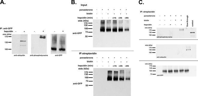
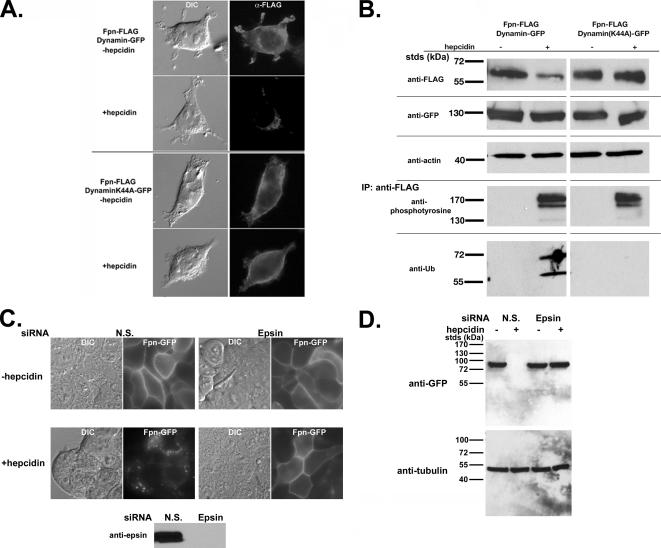
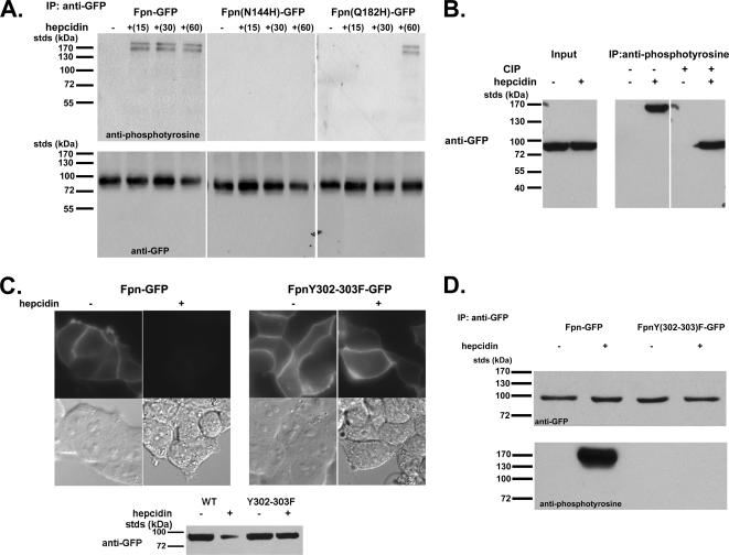
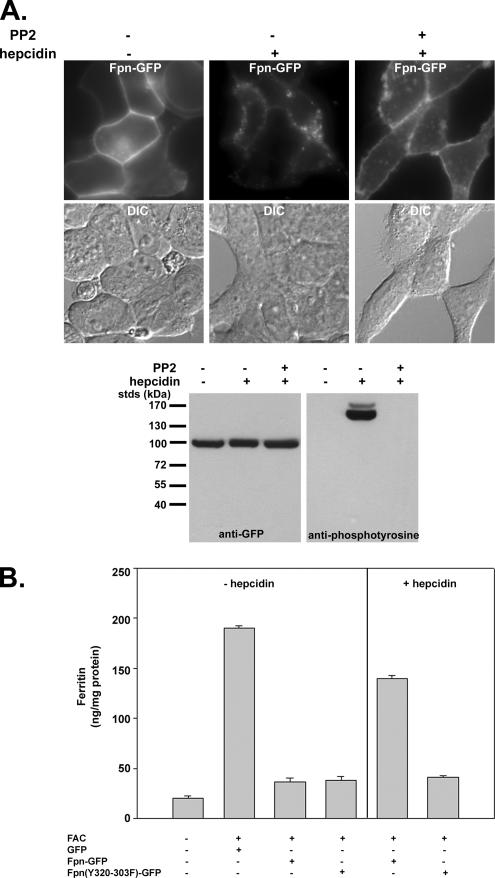
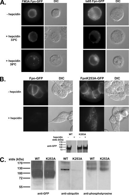
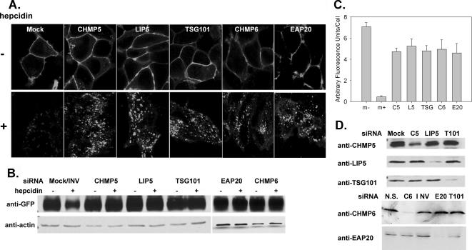
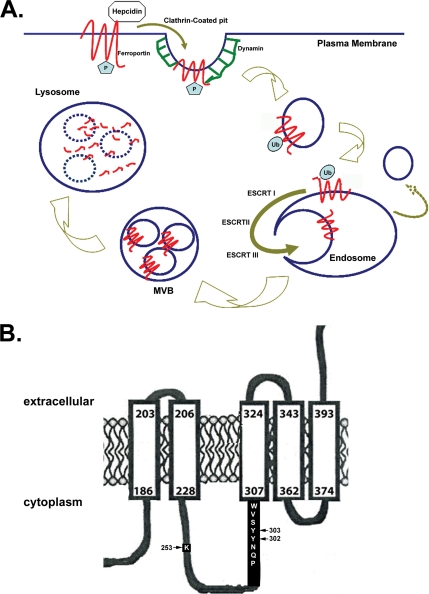
Ferroportin (Fpn) is the only known iron exporter in vertebrates. Hepcidin, a peptide secreted by the liver in response to iron or inflammation, binds to Fpn, inducing its internalization and degradation. We show that after binding of hepcidin, Fpn is tyrosine phosphorylated at the plasma membrane. Mutants of human Fpn that do not get internalized or that are internalized slowly show either absent or impaired phosphorylation. We identify adjacent tyrosines as the phosphorylation sites and show that mutation of both tyrosines prevents hepcidin-mediated Fpn internalization. Once internalized, Fpn is dephosphorylated and subsequently ubiquitinated. An inability to ubiquitinate Fpn does not prevent hepcidin-induced internalization, but it inhibits the degradation of Fpn. Ubiquitinated Fpn is trafficked through the multivesicular body pathway en route to degradation in the late endosome/lysosome. Depletion of proteins involved in multivesicular body trafficking (Endosome Sorting Complex Required for Transport proteins), by small-interfering RNA, reduces the trafficking of Fpn-green fluorescent to the lysosome.


Figures

Figure 1.
Figure 1.
Fpn-GFP is ubiquitinated and phosphorylated. (A) HEK293T cells (stably transfected with an inducible Fpn-GFP) induced to express Fpn-GFP (+ponasterone) were incubated in the presence or absence of 1 μg/ml hepcidin for 15 min. Cells were placed at 0°C and solubilized in 1.0% Triton X-100, 150 mM NaCl, 10 mM EDTA, 10 mM Tris, pH 7.4, protease inhibitor mixture, 50 mM N-ethylmaleimide, and protein phosphatase inhibitor set. Samples were immunoprecipitated with rabbit anti-GFP antibodies as described in Materials and Methods. Immunoprecipitated samples were analyzed by Western blots probing for ubiquitin by using mouse anti-ubiquitin or for phosphotyrosine by using mouse anti-phosphotyrosine followed by a peroxidase-conjugated goat anti-mouse IgG as secondary antibody. Blots were also probed for Fpn-GFP by using rabbit anti-GFP followed by peroxidase-conjugated goat anti-rabbit IgG as secondary. (B) HEK293T cells were induced to express Fpn-GFP with ponasterone for 18 h. Cells were incubated in the presence or absence of 1 μg/ml hepcidin for the indicated times, placed at 0°C, and the cell surface was biotinylated using sulfo-NHS-SS-biotin. After biotinylation, cells were solubilized as described in A, and the biotinylated proteins were affinity purified using streptavidin affinity gel. The affinity-purified samples were analyzed by Western blot using a rabbit anti-GFP followed by peroxidase-conjugated goat anti-rabbit IgG. (C) HEK293T Fpn-GFP cells were induced to express Fpn-GFP as described in B. Cells were incubated in the presence or absence of 1 μg/ml hepcidin for 15 min, placed at 0°C, and the cell surface was biotinylated. Biotinylated samples were purified using streptavidin beads as described in B. Affinity-purified samples were analyzed by Western blot with mouse anti-phosphotyrosine, mouse anti-ubiquitin followed by a peroxidase-conjugated goat anti-mouse IgG as secondary, or Fpn-GFP by using rabbit anti-GFP followed by peroxidase-conjugated goat anti-rabbit IgG as secondary. “Control” in the top panel is a mixture of tyrosine-modified proteins purchased from Calbiochem as a control for the anti-phosphotyrosine antibody. Flow-through is the sample that did not bind the streptavidin affinity gel (nonbiotinylated Fpn-GFP) but that was subsequently immunopurified using rabbit anti-GFP and the immunopurified sample analyzed by Western blot to test for the presence of phosphotyrosine or ubiquitin modifications on Fpn-GFP.
Figure 2.
Figure 2.
Fpn is phosphorylated at the plasma membrane. (A) HEK293T cells were transiently transfected with Fpn-FLAG and either Dynamin-GFP or DynaminK44A-GFP. Cells were incubated in the presence or absence of 1 μg/ml hepcidin for 60 min and processed for immunofluorescence using mouse anti-FLAG followed by Alexa 594-conjugated goat anti-mouse IgG (1:750) as secondary antibody. (B) HEK293T cells were transiently transfected as described in A. Cells were incubated in the presence or absence of 1 μg/ml hepcidin as in A, extracts obtained as described in Figure 1 and Western blotted for Fpn-FLAG, Dynamin-GFP, and actin (loading control). Extracts were immunoprecipitated using anti-FLAG resin (M2), and the immunoprecipitates were examined for the presence of tyrosine phosphorylated Fpn-FLAG or ubiquitinated Fpn-FLAG by using mouse anti-phosphotyrosine or mouse anti-ubiquitin antibodies followed by peroxidase-conjugated goat anti-mouse IgG. (C and D) HEK293T Fpn-GFP cells were transfected with nonspecific (NS) or epsin-specific siRNA oligonucleotide pools. After 48 h of incubation, cells were induced to express Fpn-GFP and then 18 h later 1 μg/ml hepcidin was added for 1 h. The efficiency of epsin depletion was assessed by Western blot analysis using antibodies to epsin. The presence of Fpn-GFP was assessed by epifluorescence (C) and Western blot analysis (D).
Figure 3.
Figure 3.
Phosphorylation, location, and function of human Fpn mutations. (A) HEK293T cells were transiently transfected with Fpn-GFP, Fpn(N144H)-GFP, or Fpn(Q182H)-GFP expressed under a CMV promoter. Cells were incubated in the presence or absence of 1 μg/ml hepcidin for indicated times, solubilized, and immunoprecipitated with rabbit anti-GFP antibodies. Immunoprecipitated samples were analyzed on nonreducing 10% SDS-PAGE, and Western blots were probed for either phosphotyrosine by using mouse anti-phosphotyrosine followed by a peroxidase-conjugated goat anti-mouse IgG as secondary or Fpn-GFP by using rabbit anti-GFP followed by peroxidase-conjugated goat anti-rabbit IgG as secondary. (B) HEK293T cells expressing wild-type Fpn-GFP were incubated with and without 1 μg/ml hepcidin for 15 min. Cell extracts were immunoprecipitated with anti-phosphotyrosine antibodies, and the immunoprecipitate was incubated in the presence or absence of calf intestinal phosphatase (CIP) for 60 min at 37°C. The samples were analyzed by SDS-PAGE and Western blot probing for Fpn-GFP. (C) HEK293T cells transfected with Fpn-GFP or Fpn(Y302-303F)-GFP were incubated in the presence of 1 μg/ml hepcidin for up to 24 h. Fpn-GFP localization was examined by fluorescence microscopy after 4 h of hepcidin incubation, and Fpn-GFP levels were measured by Western blot analysis after 24 h of hepcidin incubation. (D) HEK293T cells transfected with Fpn-GFP or Fpn(Y302-303F)-GFP were incubated with hepcidin for 15 min. The cells were solubilized, and Fpn-GFP was immunoprecipitated and analyzed by Western blots as described in A.
Figure 4.
Figure 4.
Effect of src kinase inhibitor PP2 on hepcidin-induced phosphorylation and internalization of Fpn-GFP. (A) HEK293T Fpn-GFP–expressing cells were incubated with 100 μM PP2 for 15 min. Hepcidin was added for an additional 20 min, and Fpn-GFP localization was examined by fluorescence microscopy, and tyrosine phosphorylation was assayed as on immunoprecipitated Fpn-GFP as described in Figure 3. (B) HEK293T cells, transfected with either pEGP vector, Fpn-GFP, or Fpn(Y302-303F)-GFP, were incubated with or without ferric ammonium citrate (FAC) (10 μM Fe) for 16 h, and the amount of cell-associated ferritin was determined. Iron-loaded cells expressing Fpn-GFP or Fpn(Y302-303F)-GFP were incubated in the presence of 1 μg/ml hepcidin for 24 h, and the amount of cell-associated ferritin was determined.
Figure 5.
Figure 5.
Ubiquitination is required for Fpn-GFP trafficking to the lysosome. (A) Wild-type (FM3A) or mutant cells (ts85) were transfected with Fpn-GFP. Cells were then incubated at the permissive (33°C) or restrictive (39°C) temperature in the presence or absence of 1 μg/ml hepcidin for 60 min. Cells were examined by epifluorescence microscopy for the localization of Fpn-GFP. (B) HEK293T cells were transiently transfected with Fpn-GFP or Fpn(K253A)-GFP and grown for 12 h. Cells were incubated in the presence or absence of 1 μg/ml hepcidin for 60 min at 37°C, and then they were examined for the localization of Fpn-GFP, and protein levels were assessed by Western blot. (C) Cells expressing wild-type or Fpn(K253A)-GFP were incubated in the presence of 1 μg/ml hepcidin for 15 min. The cells were solubilized, and Fpn-GFP was immunoprecipitated as in Figure 1. Immunoprecipitates were analyzed by SDS-PAGE and Western blot for the presence of Fpn-GFP by using rabbit anti-GFP followed by peroxidase-conjugated goat anti-rabbit IgG or for ubiquitinated or tyrosine-phosphorylated Fpn-GFP by using mouse anti-ubiquitin or mouse anti-phosphotyrosine antibodies followed by peroxidase-conjugated goat anti-mouse IgG.
Figure 6.
Figure 6.
Depletion of MVB proteins affects the trafficking and degradation of Fpn-GFP. (A) HEK293T Fpn-GFP cells were transfected with siRNA oligonucleotide pools specific for TSG101 (ESCRT I), EAP20 (ESCRT II), CHMP6 (ESCRT III), CHMP5 (ESCRT III), LIP5, nonspecific (N.S.), or inverted TSG101 (INV) by using OligofectAMINE. Forty-eight hours later, cells were induced to express Fpn-GFP, and after 18 h, the cells were incubated in the presence or absence of hepcidin. Cells were examined by confocal microscopy, and the localization of Fpn-GFP was determined. (B) Cells treated as described in A were solubilized as in Figure 1, analyzed on SDS-PAGE under nonreducing conditions, and Western blotted for either Fpn-GFP or actin. (C) Images from A were analyzed using National Institutes of Health ImageJ, and the amount of relative fluorescence per cell was determined. The data are expressed as arbitrary fluorescence units per cell, and error bars represent the analysis of greater than five fields per sample. (D) To assess silencing, samples from A were applied to SDS-PAGE under reducing conditions and analyzed by Western blot by using mouse or rabbit antibodies against TSG101 (T101), EAP20 (E20), CHMP6 (C6), CHMP5 (C5), or LIP5 followed by peroxidase-conjugated goat anti-mouse/rabbit IgG.
Figure 7.
Figure 7.
Model for Fpn internalization and degradation. (A) Hepcidin binds Fpn at the plasma membrane where Fpn is tyrosine phosphorylated. Once Fpn is internalized, the phosphates are removed, and Fpn is ubiquitinated, which targets it to the MVB for degradation in the lysosomes. (B) Topology of Fpn showing the potential transmembrane domain containing the phosphorylation (Y302 and Y303) and ubiquitination (K253) sites.

References

    1. Abboud S., Haile D. J. A novel mammalian iron-regulated protein involved in intracellular iron metabolism. J. Biol. Chem. 2000;275:19906–19912. - PubMed
    1. Azmi I., Davies B., Dimaano C., Payne J., Eckert D., Babst M., Katzmann D. J. Recycling of ESCRTs by the AAA-ATPase Vps4 is regulated by a conserved VSL region in Vta1. J. Cell Biol. 2006;172:705–717. - PMC - PubMed
    1. Babst M., Katzmann D. J., Estepa-Sabal E. J., Meerloo T., Emr S. D. Escrt-III: an endosome-associated heterooligomeric protein complex required for mvb sorting. Dev. Cell. 2002a;3:271–282. - PubMed
    1. Babst M., Katzmann D. J., Snyder W. B., Wendland B., Emr S. D. Endosome-associated complex, ESCRT-II, recruits transport machinery for protein sorting at the multivesicular body. Dev. Cell. 2002b;3:283–289. - PubMed
    1. Babst M., Odorizzi G., Estepa E. J., Emr S. D. Mammalian tumor susceptibility gene 101 (TSG101) and the yeast homologue, Vps23p, both function in late endosomal trafficking. Traffic. 2000;1:248–258. - PubMed

Publication types

MeSH terms