

Differential encoding of losses and gains in the human striatum
- PMID: 17475790
- PMCID: PMC2630024
- DOI: 10.1523/JNEUROSCI.0400-07.2007
Differential encoding of losses and gains in the human striatum
Abstract
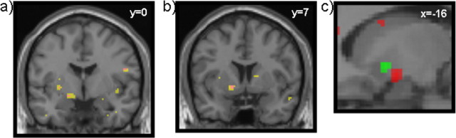
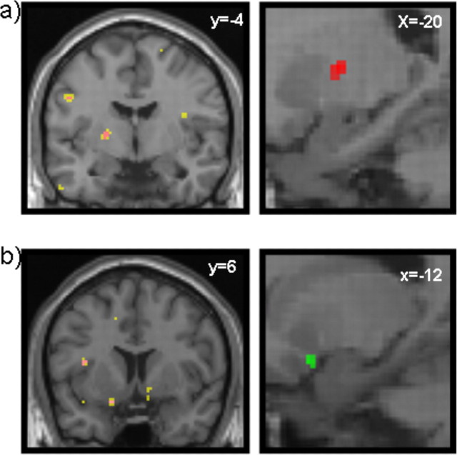
Studies on human monetary prediction and decision making emphasize the role of the striatum in encoding prediction errors for financial reward. However, less is known about how the brain encodes financial loss. Using Pavlovian conditioning of visual cues to outcomes that simultaneously incorporate the chance of financial reward and loss, we show that striatal activation reflects positively signed prediction errors for both. Furthermore, we show functional segregation within the striatum, with more anterior regions showing relative selectivity for rewards and more posterior regions for losses. These findings mirror the anteroposterior valence-specific gradient reported in rodents and endorse the role of the striatum in aversive motivational learning about financial losses, illustrating functional and anatomical consistencies with primary aversive outcomes such as pain.
Figures
Similar articles
-
Striatal and Pallidal Activation during Reward Modulated Movement Using a Translational Paradigm.J Int Neuropsychol Soc. 2015 Jul;21(6):399-411. doi: 10.1017/S1355617715000491. Epub 2015 Jul 9. J Int Neuropsychol Soc. 2015. PMID: 26156687 Free PMC article.
-
Overlapping prediction errors in dorsal striatum during instrumental learning with juice and money reward in the human brain.J Neurophysiol. 2009 Dec;102(6):3384-91. doi: 10.1152/jn.91195.2008. Epub 2009 Sep 30. J Neurophysiol. 2009. PMID: 19793875
-
Reduced striatal responses to reward prediction errors in older compared with younger adults.J Neurosci. 2013 Jun 12;33(24):9905-12. doi: 10.1523/JNEUROSCI.2942-12.2013. J Neurosci. 2013. PMID: 23761885 Free PMC article.
-
The role of the striatum in aversive learning and aversive prediction errors.Philos Trans R Soc Lond B Biol Sci. 2008 Dec 12;363(1511):3787-800. doi: 10.1098/rstb.2008.0161. Philos Trans R Soc Lond B Biol Sci. 2008. PMID: 18829426 Free PMC article. Review.
-
Prediction error in reinforcement learning: a meta-analysis of neuroimaging studies.Neurosci Biobehav Rev. 2013 Aug;37(7):1297-310. doi: 10.1016/j.neubiorev.2013.03.023. Epub 2013 Apr 6. Neurosci Biobehav Rev. 2013. PMID: 23567522 Review.
Cited by
-
The human insula processes both modality-independent and pain-selective learning signals.PLoS Biol. 2022 May 6;20(5):e3001540. doi: 10.1371/journal.pbio.3001540. eCollection 2022 May. PLoS Biol. 2022. PMID: 35522696 Free PMC article.
-
Positive and negative reinforcement activate human auditory cortex.Front Hum Neurosci. 2013 Dec 5;7:842. doi: 10.3389/fnhum.2013.00842. eCollection 2013. Front Hum Neurosci. 2013. PMID: 24367318 Free PMC article.
-
Do Adolescents Use Substances to Relieve Uncomfortable Sensations? A Preliminary Examination of Negative Reinforcement among Adolescent Cannabis and Alcohol Users.Brain Sci. 2020 Apr 5;10(4):214. doi: 10.3390/brainsci10040214. Brain Sci. 2020. PMID: 32260480 Free PMC article.
-
Differential reinforcement learning responses to positive and negative information in unmedicated individuals with depression.Eur Neuropsychopharmacol. 2021 Dec;53:89-100. doi: 10.1016/j.euroneuro.2021.08.002. Epub 2021 Sep 10. Eur Neuropsychopharmacol. 2021. PMID: 34517334 Free PMC article.
-
Levodopa administration modulates striatal processing of punishment-associated items in healthy participants.Psychopharmacology (Berl). 2015 Jan;232(1):135-44. doi: 10.1007/s00213-014-3646-7. Epub 2014 Jun 13. Psychopharmacology (Berl). 2015. PMID: 24923987 Free PMC article. Clinical Trial.
References
-
- Barto AG. Adaptive critic and the basal ganglia. In: Houk JC, Davis JL, Beiser DG, editors. Models of information processing in the basal ganglia. Cambridge: MIT; 1995. pp. 215–232.
-
- Becerra L, Breiter HC, Wise R, Gonzalez RG, Borsook D. Reward circuitry activation by noxious thermal stimuli. Neuron. 2001;32:927–946. - PubMed
-
- Bitsios P, Szabadi E, Bradshaw CM. The fear-inhibited light reflex: importance of the anticipation of an aversive event. Int J Psychophysiol. 2004;52:87–95. - PubMed
-
- Breiter HC, Aharon I, Kahneman D, Dale A, Shizgal P. Functional imaging of neural responses to expectancy and experience of monetary gains and losses. Neuron. 2001;30:619–639. - PubMed
Publication types
MeSH terms
Grants and funding
LinkOut - more resources
Full Text Sources