Clinical Trial
. 2007 Sep 1;110(5):1458-65.
doi: 10.1182/blood-2007-02-075184. Epub 2007 May 2.

The human Shwachman-Diamond syndrome protein, SBDS, associates with ribosomal RNA

Affiliations
Clinical Trial

The human Shwachman-Diamond syndrome protein, SBDS, associates with ribosomal RNA

Karthik A Ganapathi et al. Blood. .

Abstract

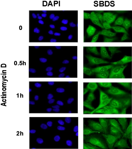
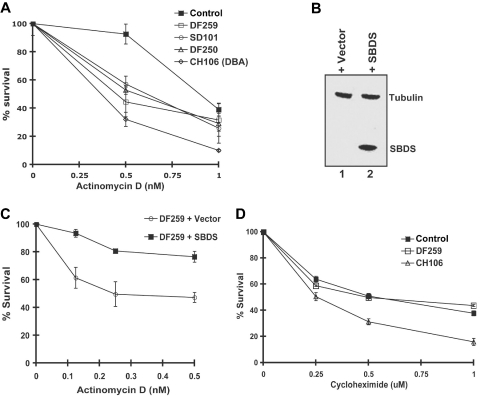
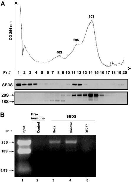
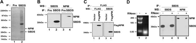
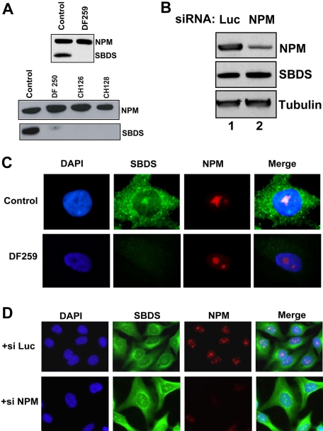
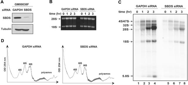
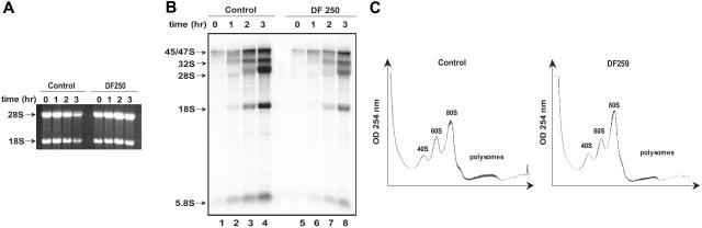
Shwachman-Diamond syndrome (SDS) is an autosomal recessive disorder characterized by bone marrow failure, exocrine pancreatic dysfunction, and leukemia predisposition. Mutations in the SBDS gene are identified in most patients with SDS. SBDS encodes a highly conserved protein of unknown function. Data from SBDS orthologs suggest that SBDS may play a role in ribosome biogenesis or RNA processing. Human SBDS is enriched in the nucleolus, the major cellular site of ribosome biogenesis. Here we report that SBDS nucleolar localization is dependent on active rRNA transcription. Cells from patients with SDS or Diamond-Blackfan anemia are hypersensitive to low doses of actinomycin D, an inhibitor of rRNA transcription. The addition of wild-type SBDS complements the actinomycin D hypersensitivity of SDS patient cells. SBDS migrates together with the 60S large ribosomal subunit in sucrose gradients and coprecipitates with 28S ribosomal RNA (rRNA). Loss of SBDS is not associated with a discrete block in rRNA maturation or with decreased levels of the 60S ribosomal subunit. SBDS forms a protein complex with nucleophosmin, a multifunctional protein implicated in ribosome biogenesis and leukemogenesis. Our studies support the addition of SDS to the growing list of human bone marrow failure syndromes involving the ribosome.


Figures

Figure 1
Figure 1
Nucleolar localization of SBDS is abrogated by actinomycin D treatment. HeLa cells were treated with 2 nM actinomycin D for the times indicated. Cells were fixed and stained for SBDS (green) and counterstained with DAPI (blue) to visualize nuclei (63× magnification). See “Patients, materials, and methods; Immunofluoresence” for image acquisition details.
Figure 2
Figure 2
Cells from SDS patients are hypersensitive to actinomycin D in an SBDS-dependent manner. (A) Lymphoblasts from a healthy control, SDS patients (DF250, DF259, and SD101), and an RPS19+ DBA patient cell (CH106) were plated in the presence of increasing concentrations of actinomycin D. Cell viability was assayed after 72 hours. Assays were performed in triplicate per experiment and repeated for a minimum of 3 independent experiments. Bars represent the standard error. (B) DF259 cells were infected with lentivirus containing GFP alone or together with wild-type SBDS cDNA downstream of an IRES sequence. Lentiviral infection was greater than 90% as observed by fluorescence microscopy. Cell lysates were analyzed by immunoblotting with antibodies against SBDS and tubulin. (C) DF259 cells infected with lentivirus containing empty vector or full-length SBDS cDNA were plated in the presence of increasing concentrations of actinomycin D. Cell viability was assayed in triplicate per experiment and repeated for 3 independent experiments. Bars represent the standard error. (D) Lymphoblasts (normal control, DF259, and CH106) were plated in the presence of increasing concentrations of cycloheximide. Cell viability was assayed after 72 hours in triplicate for each experiment for a total of 3 independent experiments. Bars represent the standard error.
Figure 3
Figure 3
SBDS cosediments with the 60S ribosomal precursor subunit and associates with the 28S ribosomal RNA. (A) HeLa cell lysates were fractionated on a 10% to 30% sucrose gradient by ultracentrifugation. Absorbance at 254 nM across the gradient is shown (top panel). Proteins were precipitated from equal aliquots of each fraction and immunoblotted for SBDS (middle panel). RNA was extracted from equal volumes of each fraction and analyzed by agarose gel electrophoresis and ethidium bromide staining (bottom panel). Data shown are representative of results obtained from 3 independent experiments. (B) Endogenous SBDS was immunoprecipitated from nuclear extracts of HeLa cells, healthy control lymphoblasts (control), or DF277 lymphoblasts derived from an SDS patient. Preimmune serum was used as a negative control for the immunoprecipitation (lane 2). RNA was extracted from the immunoprecipitates and analyzed by agarose gel electrophoresis and ethidium bromide staining. Data shown are representative of results obtained from 3 independent experiments.
Figure 4
Figure 4
SBDS associates with nucleophosmin in an RNA-independent manner. (A) Endogenous SBDS was immunoprecipitated from HeLa nuclear extracts with an anti-SBDS antibody. The immunoprecipitated proteins were separated by sodium dodecyl sulfate–polyacrylamide gel electrophoresis on a 4% to 12% Bis-Tris gel followed by Coomassie blue staining. Preimmune serum (Pre) was used as a negative control for the immunoprecipitation (lane 1). Protein bands from the SBDS immunoprecipitate (lane 2) were excised and analyzed by tryptic digestion and matrix-assisted laser description/ionisation time of flight (MALDI-TOF) mass spectrometry. (B) HeLa cell lysates were incubated with either anti-SBDS antibody or preimmune serum (Pre). The immunoprecipitates were analyzed by Western blotting for SBDS (lanes 1 and 2) and NPM (lanes 3 and 4). (C) 293T cells were transfected with either FLAG-NPM (lanes 2 and 4) or empty pFLAG-CMV vector (lanes 1 and 3). Cell lysates were immunoprecipitated with an anti-FLAG antibody. The resulting precipitates were immunoblotted for FLAG (lanes 1 and 2) or SBDS (lanes 3 and 4). (D) HeLa cell lysates were incubated at 30°C for 20 minutes in the presence (lane 2) or absence (lane 1) of 4 μg RNase A. RNA was extracted and analyzed by agarose gel electrophoresis and ethidium bromide staining (left panel, lanes 1 and 2). SBDS was immunoprecipitated from the mock-treated and RNase-treated lysates. The resulting pellets were analyzed by immunoblotting (right panel) for SBDS (lanes 3 and 4) and NPM (lanes 5 and 6).
Figure 5
Figure 5
SBDS is not required for NPM protein stability or nucleolar localization. (A) Primary fibroblast lysates from healthy controls or SDS patients (DF259, DF250, CH126, CH128) were immunoblotted for SBDS and NPM. (B) HeLa cells were transfected with siRNA against either luciferase (Luc) or NPM. Cells were lysed 72 hours after transfection and immunoblotted for SBDS, NPM, and tubulin. (C) Normal control and DF259 primary fibroblasts were fixed and stained for SBDS (green) and NPM (red) and counterstained with DAPI (blue) to visualize the nuclei. (D) HeLa cells were transfected with siRNA against either luciferase (++ si Luc) or NPM (+ si NPM) for 48 hours. Cells were fixed and stained for SBDS (green) and NPM (red) and counterstained with DAPI (blue). See “Patients, materials, and methods; Immunofluorescence” for image acquisition details.
Figure 6
Figure 6
SBDS loss is not associated with a discrete block in rRNA processing. (A) siRNA against either GAPDH or SBDS was introduced into human skin fibroblasts (GM00038). Cells were lysed 72 hours later and immunoblotted for SBDS and tubulin. GAPDH knock down was confirmed to be greater than 50% by immunoblot densitometry and quantitative PCR (data not shown). (B) GM00038 fibroblasts containing siRNA against GAPDH or SBDS from (A) were metabolically labeled with 32P-orthophosphate for 75 minutes and chased with 25 mM phosphate for the indicated times in hours. RNA was extracted at the indicated time points, resolved on a 1% agarose/formaldehyde gel, and stained with ethidium bromide to confirm equal loading. (C) RNA from the gel in panel B was transferred to a nylon membrane and analyzed by autoradiography. The positions of the 45S/47S and 32S precursor rRNAs and of the mature 28S, 18S, and 5.8S rRNAs are indicated. (D) GM00038 fibroblasts containing siRNA against GAPDH or SBDS were lysed and fractionated on 10% to 45% sucrose gradients by ultracentrifugation. Absorbance at 254 nM was measured across the gradient, and the positions corresponding to the 40S, 60S, and 80S ribosomal particles are indicated. Results shown are representative of 3 independent experiments.
Figure 7
Figure 7
SDS patient cell lines show decreased ribosomal RNA synthesis. (A) Healthy control and SDS patient (DF250) primary fibroblasts were metabolically labeled with 32P-orthophosphate for 75 minutes and chased with 25 mM phosphate for the indicated times in hours. RNA was extracted at the indicated time points, resolved on a 1% agarose/formaldehyde gel, and stained with ethidium bromide. (B) RNA from the gel in panel A was transferred to a nylon membrane and analyzed by autoradiography. The positions of the 45S and 32S rRNA precursor rRNAs and of the mature 28S, 18S, and 5.8S rRNAs are indicated. (C) Lysates from normal control primary fibroblasts or SDS patient primary fibroblasts were sedimented through sucrose gradients as in Figure 6D. This experiment was repeated for a total of 3 independent experiments. No consistent difference in the ratios of the 40S, 60S, and 80S peaks was noted.

References

    1. Shwachman H, Diamond LK, Oski FA, Khaw KT. The syndrome of pancreatic insufficiency and bone marrow dysfunction. J Pediatr. 1964;65:645–663. - PubMed
    1. Bodian M, Sheldon W, Lightwood R. Congenital hypoplasia of the exocrine pancreas. Acta Paediatr. 1964;53:282–293. - PubMed
    1. Shimamura A. Shwachman-Diamond syndrome. Semin Hematol. 2006;43:178–188. - PubMed
    1. Dror Y. Shwachman-Diamond syndrome. Pediatr Blood Cancer. 2005;45:892–901. - PubMed
    1. Boocock GR, Morrison JA, Popovic M, et al. Mutations in SBDS are associated with Shwachman-Diamond syndrome. Nat Genet. 2003;33:97–101. - PubMed

Publication types

MeSH terms