. 2007 May 2;2(5):e416.
doi: 10.1371/journal.pone.0000416.

Short-term exposure of multipotent stromal cells to low oxygen increases their expression of CX3CR1 and CXCR4 and their engraftment in vivo

Affiliations

Short-term exposure of multipotent stromal cells to low oxygen increases their expression of CX3CR1 and CXCR4 and their engraftment in vivo

Shih-Chieh Hung et al. PLoS One. .

Abstract

The ability of stem/progenitor cells to migrate and engraft into host tissues is key to their potential use in gene and cell therapy. Among the cells of interest are the adherent cells from bone marrow, referred to as mesenchymal stem cells or multipotent stromal cells (MSC). Since the bone marrow environment is hypoxic, with oxygen tensions ranging from 1% to 7%, we decided to test whether hypoxia can upregulate chemokine receptors and enhance the ability of human MSCs to engraft in vivo. Short-term exposure of MSCs to 1% oxygen increased expression of the chemokine receptors CX3CR1and CXCR4, both as mRNA and as protein. After 1-day exposure to low oxygen, MSCs increased in vitro migration in response to the fractalkine and SDF-1alpha in a dose dependent manner. Blocking antibodies for the chemokine receptors significantly decreased the migration. Xenotypic grafting into early chick embryos demonstrated cells from hypoxic cultures engrafted more efficiently than cells from normoxic cultures and generated a variety of cell types in host tissues. The results suggest that short-term culture of MSCs under hypoxic conditions may provide a general method of enhancing their engraftment in vivo into a variety of tissues.


Conflict of interest statement

Competing Interests: The authors have declared that no competing interests exist.

Figures

Figure 1
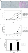
Figure 1. Effect of oxygen tensions and initial plating densities on apoptosis and expansion of MSCs.
(A) The MSCs at passage 3 were subjected to normoxic (Nor), hypoxic (Hyp) or serum free conditions (SF) for 2 days. Apoptosis was evaluated by the APOPercentage assay and the apoptotic cells are stained as pink. (B,C) Passage 3 MSCs were plated on 60-cm2 dishes at 50 and 1,000 cells/cm2 and cultured in normoxic and hypoxic conditions. The cells were harvested and counted up to 10 days. (B) Total cell numbers per 60-cm2 dish and (C) fold increase are shown. Data are expressed as mean±standard deviation (n = 3). (D) Passage 3 MSCs were plated at 100 cells/60-cm2 dish, cultured in normoxic or hypoxic conditions for 10 or 14 days, and stained with Crystal Violet to give total colony count. Colony numbers, which indicate colony-forming efficiency (%), are shown. Data are expressed as mean±standard deviation (n = 3). (*, p<0.05, Student's t test).
Figure 2
Figure 2. Hypoxia inhibits osteogenesis and adipogenesis.
The MSCs were plated at 100 cells/60-cm2 dish, cultured in normoxic (Nor) or hypoxic (Hyp) conditions for 7 days, and replaced with osteogenic or adipogenic induction media for additional 7 to 21 days. (A) Total colonies stained with Crystal Violet (upper two panels) and osteogenic colonies stained with Alizarin Red-S (lower two panels). (B) The panel shows the numbers of Alizarin Red-S and Oil Red-O-positive colonies after culture in osteogenic and adipogenic induction media, respectively. Data are expressed as mean±standard deviation (n = 3). (*, p<0.05, Student's t test). (C) Cells were plated at 10,000 cells/cm2, and then cultured in adipogenic induction media under normoxic or hypoxic conditions. Oil Red-O staining was performed at 2 weeks after induction to visualize the level of fat production in the cells (red stain).
Figure 3
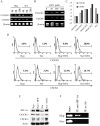
Figure 3. Effect of hypoxia on CX3CR1 and CXCR4 expression by early culture of MSCs.
(A) The MSCs were cultured for 4–20 h in normoxic (Nor) or hypoxic (Hyp) conditions, as indicated. Total RNA was analyzed by RT-PCR for CX3CR1, CX3CR4, and b-actin mRNA expression. (B) Total RNA from MSCs cultured for 20 h in the presence of increasing concentrations of DFX was analyzed by RT-PCR for CX3CR1 and CXCR4 mRNA expression. (C) Quantitaive RT-PCR for CXCR4 and CX3CR1 mRNA expression at 20 h of treatment. The vertical axis represents the relative ratio of indicated condition to normoxic condition without the addition of DFX.. (D) Surface expression of CXCR4 and CX3CR1 were determined by flow cytometry using a mouse mAb anti-CXCR4 and a rat mAb anti-CX3CR1, respectively (continuous line). Irrelevant antibody is indicated by the dotted line. The results are representative of three independent experiments. (E) Hypoxia and DFX-treated MSCs were cultured for 24 h in the indicated conditions and analyzed by western blot for HIF-1α, CXCR4, CX3CR1, and actin protein levels. (F) HIF-1α binding to the CX3CR1 promoter. The MSCs were cultured for 4 h in normoxic or hypoxic condition. ChIP was performed with or without rabbit antibody specific for HIF-1α. There is no difference between PCR of the CX3CR1 promoter using input chromatin for normoxic and hypoxic cells. However, a band was noted for hypoxic cells but not for normoxic cells, when PCR of the CX3CR1 promoter was performed using immunoprecipitation with HIF-1α antibody.
Figure 4
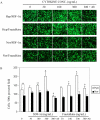
Figure 4. Effect of hypoxia on the chemotactic response of MSCs to SDF-1a and fractalkine.
(A) Cells were cultured for 22 h in normoxic (Nor) or hypoxic (Hyp) conditions. Migration of MSCs was assayed by chemotaxis microchamber technique. Effect of hypoxia on the chemotactic response of MSCs to increasing concentrations of SDF-1a (0–300 ng/mL) and fractalkine (0–300 ng/mL) was assessed by counting the total number of cells in 5 power fields. For inhibition studies of MSCs migration, MSCs were preincubated with 10 mg/mL of anti-CXCR4 or fractalkine-containing medium was preincubated with 10 mg/mL of anti-fractalkine. (B) Results are mean±SD of five power fields. (*, p<0.01, **, p<0.05 as compared with control without cytokine, #, p<0.01 as compared with that with cytokine added at 300 ng/mL, Student's t test).
Figure 5
Figure 5. Dye-labeled MSCs transplanted in 2-day-old chicken embryo and detected in organs of 5-day-old chicken embryo by epifluorescence microscopy.
(A) Examples of hypoxia-exposed/CMFDA-labeled (green) and normoxic condition-exposed/CMTMR cells (red) in the heart, brain or spine area. 4',6-Diamidino-2-phenylindole (DAPI) stain was used to identify nuclei (mag: 400×). (B) Cells per 400×powered field. Results are mean±SD of more than 20 power fields from different embryos. (*, p<0.05, Student's t test).
Figure 6
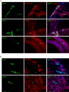
Figure 6. Epifluorescent immunohistology of sections from chick embryos infused with GFP+MSCs.
(A) Cells (arrows) positive for GFP and cardiotin in heart (upper row); cells (arrows) positive for GFP and a-myosin heavy chain (MHC) in heart (middle row); cells (arrows) positive for GFP and NF-H in brain (bottom row). (B) Cells (arrows) positive for GFP, but negative for a chicken-specific marker (CSM) in heart (upper row) or brain area (lower row). DAPI stain was used to identify nuclei (mag: 400×).

References

    1. Prockop DJ, Gregory CA, Spees JL. One strategy for cell and gene therapy: harnessing the power of adult stem cells to repair tissues. Proc Natl Acad Sci U S A. 2003;(100 Suppl 1):11917–11923. - PMC - PubMed
    1. Jiang Y, Jahagirdar BN, Reinhardt RL, Schwartz RE, Keene CD, et al. Pluripotency of mesenchymal stem cells derived from adult marrow. Nature. 2002;418:41–49. - PubMed
    1. Matsuzaki Y, Kinjo K, Mulligan RC, Okano H. Unexpectedly efficient homing capacity of purified murine hematopoietic stem cells. Immunity. 2004;20:87–93. - PubMed
    1. Owen M, Friedenstein AJ. Stromal stem cells: marrow-derived osteogenic precursors. Ciba Found Symp. 1988;136:42–60. - PubMed
    1. Chou SH, Kuo TK, Liu M, Lee OK. In utero transplantation of human bone marrow-derived multipotent mesenchymal stem cells in mice. J Orthop Res. 2006;24:301–312. - PubMed

Publication types