Review
. 2007 May;117(5):1195-203.
doi: 10.1172/JCI31205.

DC-based cancer vaccines

Affiliations
Review

DC-based cancer vaccines

Eli Gilboa. J Clin Invest. 2007 May.

Abstract

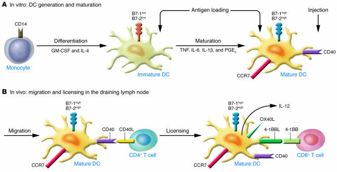
Because of the large preexisting antigenic load and immunosuppressive environment within a tumor, inducing therapeutically useful antitumor immunity in cancer patients requires the development of powerful vaccination protocols. An approach gaining increasing popularity in the tumor vaccine field is to immunize cancer patients with their own DCs loaded ex vivo with tumor antigens. The underlying premise of this approach is that the efficiency and control over the vaccination process provided by ex vivo manipulation of the DCs generates an optimally potent APC and a superior method for stimulating antitumor immunity in vivo compared with the more conventional direct vaccination methods, offsetting the added cost and complexity associated with this form of customized cell therapy.


Figures

Figure 1
Figure 1. Ex vivo differentiation and activation of DCs for cancer immunotherapy.
(A) The most common method used to generate DCs for clinical trials is to culture CD14+ monocytes in serum-free media in the presence of GM-CSF and IL-4. Following 5–7 days in culture, the monocytes differentiate into immature DCs, which lose CD14 expression and express moderate to low levels of CD40 and the costimulatory ligands B7-1 and B7-2. DC maturation is accomplished by culturing the immature DCs for an additional 24–48 hours in the presence of several biological agents, the most popular combination being TNF, IL-6, IL-1β, and PGE2 (41). Mature DCs further upregulate CD40, B7-1, and B7-2 and induce the de novo expression of the lymph node homing receptor CC chemokine receptor 7 (CCR7). Antigen loading occurs at either the immature or mature DC stage. (B) Mature antigen-loaded DCs are injected into patients subcutaneously, intradermally, or intravenously. They migrate to the draining lymph node, where they encounter and present antigen (not shown) to cognate CD4+ T cells. Cross-linking CD40 on the DCs by CD40L, which is expressed on the antigen-activated CD4+ T cell, induces the mature DCs to differentiate further, a process known as licensing. Licensed DCs upregulate additional cell surface products, notably the ligands for OX40 and 4-1BB (OX40L and 4-1BBL, respectively). The licensed DCs present antigen to cognate CD8+ T cells. 4-1BBL–mediated costimulation through 4-1BB on the antigen-activated CD8+ T cells enhances the survival and proliferative capacity of the activated CD8+ T cells. Likewise, OX40L-mediated costimulation enhances the survival and proliferation of the activated CD4+ T cells (not shown).
Figure 2
Figure 2. The MHC class I and class II antigen presentation pathways.
(A) The endogenous MHC class I presentation pathway leads to the activation of CD8+ T cells. Antigens expressed in the cells or introduced into the cytoplasm are degraded by the proteasome complex to generate short peptides that are translocated into the ER through special pores controlled by the transporter associated with antigen processing (TAP) proteins. In the lumen of the ER, the peptides associate with newly synthesized MHC class I molecules and the peptide–MHC class I complex is transported to the cell surface, where it is presented to CD8+ T cells expressing the cognate TCR (that is, the TCR that recognizes the particular peptide-MHC complex). Therefore, delivery into DCs of nucleic acid–encoded antigen, which needs to be translated in the cytoplasm, favors the generation of MHC class I–restricted CD8+ T cell responses. TGN, trans-Golgi network. (B) The exogenous MHC class II presentation pathway leads to the activation of CD4+ T cells. Antigens captured by professional APCs such as macrophages and DCs are routed through the endosome, where they undergo partial proteolytic degradation to generate peptides that associate with nascent MHC class II molecules. The peptide–MHC class II complexes are transported to the cell surface and presented to cognate CD4+ T cells. Therefore, generation of CD4+ T cell responses is favored by vaccination with whole protein–based antigens.

References

    1. Banchereau J., Steinman R.M. Dendritic cells and the control of immunity. Nature. 1998;392:245–252. - PubMed
    1. Banchereau J., et al. Immunobiology of dendritic cells. Annu. Rev. Immunol. 2000;18:767–811. - PubMed
    1. Reis e Sousa C. Dendritic cells as sensors of infection. Immunity. 2001;14:495–498. - PubMed
    1. Livingston P.O. Experimental and clinical studies with active specific immunotherapy. Prog. Clin. Biol. Res. 1989;288:309–321. - PubMed
    1. Gilboa E. The promise of cancer vaccines. Nat. Rev. Cancer. 2004;4:401–411. - PubMed

MeSH terms