. 2007 May;117(5):1249-59.
doi: 10.1172/JCI29710.

Diabetic impairments in NO-mediated endothelial progenitor cell mobilization and homing are reversed by hyperoxia and SDF-1 alpha

Affiliations

Diabetic impairments in NO-mediated endothelial progenitor cell mobilization and homing are reversed by hyperoxia and SDF-1 alpha

Katherine A Gallagher et al. J Clin Invest. 2007 May.

Abstract

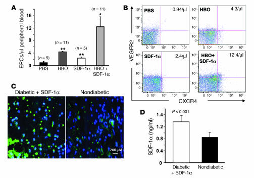
Endothelial progenitor cells (EPCs) are essential in vasculogenesis and wound healing, but their circulating and wound level numbers are decreased in diabetes. This study aimed to determine mechanisms responsible for the diabetic defect in circulating and wound EPCs. Since mobilization of BM EPCs occurs via eNOS activation, we hypothesized that eNOS activation is impaired in diabetes, which results in reduced EPC mobilization. Since hyperoxia activates NOS in other tissues, we investigated whether hyperoxia restores EPC mobilization in diabetic mice through BM NOS activation. Additionally, we studied the hypothesis that impaired EPC homing in diabetes is due to decreased wound level stromal cell-derived factor-1alpha (SDF-1alpha), a chemokine that mediates EPC recruitment in ischemia. Diabetic mice showed impaired phosphorylation of BM eNOS, decreased circulating EPCs, and diminished SDF-1alpha expression in cutaneous wounds. Hyperoxia increased BM NO and circulating EPCs, effects inhibited by the NOS inhibitor N-nitro-L-arginine-methyl ester. Administration of SDF-1alpha into wounds reversed the EPC homing impairment and, with hyperoxia, synergistically enhanced EPC mobilization, homing, and wound healing. Thus, hyperoxia reversed the diabetic defect in EPC mobilization, and SDF-1alpha reversed the diabetic defect in EPC homing. The targets identified, which we believe to be novel, can significantly advance the field of diabetic wound healing.


Figures

Figure 1
Figure 1. Impaired phosphorylation of BM eNOS with attenuation of HBO-induced NO levels results in decreased circulating EPCs in diabetic mice.
(A) Representative Western blot analysis for BM eNOS. Diabetic mice demonstrated decreased phosphorylated eNOS compared with nondiabetic controls. Insulin failed to restore impaired eNOS phosphorylation. Quantification of phospho-eNOS (p-eNOS). Results are based on 4 experiments and show the amount of phospho-eNOS relative to total eNOS and β-actin. Nondiabetic controls are used as the standard (value set at 100). *P < 0.01. (B) Changes in cell composition in BM of diabetic mice. EPC (VEGFR2/CXCR4) and HSC (CD34/CD45) populations are unchanged, while mesenchymal stromal (CD73/CD44) and inflammatory cell (SSC/CD3/CD45RA) populations are slightly decreased in diabetic mice compared with nondiabetic mice. Percentages indicate positive cells in total BM cells counted. (C) Quantification of the number of Tie2+/VEGFR2+ EPCs/μl of peripheral blood in Tie2-GFP mice by flow cytometry at 7 days following STZ treatment. A substantial reduction in circulating EPCs was found in diabetic compared with nondiabetic mice. (D and E) NO production in the BM cavity of diabetic and nondiabetic mice during 10 minutes of HBO treatment. Baseline NO levels were obtained 5 minutes prior to onset of pressurization at 100% O2 (gray bar). Solid lines represent mean values, with surrounding gray or black shading representing SEM. (D) Hyperoxia increases BM NO levels significantly, but the NO response is attenuated in diabetic mice compared with nondiabetic animals (P < 0.05). Insulin did not reverse the impairment of NO production. (E) Total iNOS and nNOS proteins were upregulated in diabetic mice. –, nondiabetic mice; +, diabetic mice. (F) Complete inhibition of BM NO production in diabetic and nondiabetic mice undergoing HBO treatment after pretreatment with l-NAME.
Figure 2
Figure 2. NO-dependent EPC, not lymphocyte, mobilization is enhanced by hyperoxia.
Flow cytometry quantification of circulating peripheral blood EPCs (CXCR4+/VEGFR2+) (A) and lymphocytes (B) in diabetic FVB and eNOS–/– mice and peripheral blood EPCs (Tie2+/VEGFR2+) in Tie2-GFP (H) mice. Data are based on 6 (A and B) and 12 (H) experiments. Mice were treated with or without HBO (HBO+ and HBO, respectively) or with l-NAME ± HBO. *P < 0.05; **P < 0.005. (CG and IP) HBO significantly increased circulating EPCs, while l-NAME inhibited this effect. Representative dot plots with number of circulating EPCs in peripheral blood of diabetic FVB (CG) and nondiabetic (IL) and diabetic (MP) Tie2-GFP mice are shown. (C, I, and M) HBO, (D, J, and N) HBO+, (E, K, and O) l-NAME, and (F, L, and P) l-NAME+HBO. (G) Isotype control (VEGFR2/CXCR4).
Figure 3
Figure 3. Decreased SDF-1α expression in peripheral wounds of diabetic mice.
(A) Quantitative detection of the SDF-1α mRNA at various time points following STZ treatment in wound tissue of diabetic and nondiabetic mice by real-time RT-PCR. SDF-1α mRNA decreased significantly at day 9. Data are based on 3 experiments. (B) Left: Epithelial cells and myofibroblasts are impaired in the production of SDF-1α in the wound tissues. Double staining (yellow) of SDF-1α (red) and cell type–specific marker (green) demonstrated a downregulated expression of SDF-1α in epithelial cells and myofibroblasts in diabetic wounds. Right: Percentage of double-positive cells per field. Data are derived from 10 sections of 3 individual experiments.
Figure 4
Figure 4. Synergistic enhancement of EPC mobilization by HBO and SDF-1α in a murine diabetic model.
(A) Diabetic mice were divided into 4 groups that received daily wound injections with either SDF-1α or PBS. Half of the mice also received daily HBO. Forty-eight hours after wounding, peripheral blood was analyzed by flow cytometry. Quantification of EPCs was performed for each group. Data are based on 10 experiments. SDF-1α+HBO–treated mice had a significant increase in circulating EPCs compared with other groups (*P < 0.05). SDF-1α- and PBS+HBO–treated groups demonstrated a statistically significant increase as compared with the PBS-treated group (**P < 0.05). (B) Representative dot plots are shown, with number of peripheral blood EPCs noted in each of the CXCR4+/VEGFR2+ quadrants. (C) Immunostaining demonstrated a supraphysiologic level of SDF-1α in diabetic wounds after SDF-1α injection compared with nondiabetic wounds. (D) Local administration of SDF-1α results in supraphysiologic systemic peripheral blood SDF-1α level above that present in nondiabetic mice. ELISA demonstrated an increased systemic SDF-1α concentration 2 hours following local wound injection with SDF-1α.
Figure 5
Figure 5. Impaired EPC homing to wound tissue in diabetes is reversed by cutaneous administration of SDF-1α.
BM cells from GFP mice were transplanted into γ-irradiated FVB mice. Four groups of wounded diabetic chimeric mice were treated with daily wound injections of PBS, HBO, SDF-1α, or HBO+SDF-1α. After 3 days of treatment, wounds were harvested and analyzed by fluorescent immunostaining of tissue sections with anti–GFP-FITC or anti–VEGFR2-PE Abs. Nuclei were counterstained with Hoescht dye. Recruited EPCs were identified as GFP+/VEGFR2+ cells. (A) Quantification of recruited EPCs in diabetic mice. For each animal, 10 random high-power fields (HPFs, ×100) from 5 serial cross-sections were analyzed, and GFP+/VEGFR2+ cells were quantified relative to the total wound cellularity. Data are based on 3 experiments. SDF-1α+HBO–treated mice had a significant increase in the amount of recruited EPCs compared with other groups (*P < 0.05). SDF-1α–treated animals had a significant increase in amount of tissue EPCs compared with PBS control (**P < 0.05). HBO did not significantly enhance EPC homing to wounds. (B) Representative fluorescent immunostaining of wound sections are shown.
Figure 6
Figure 6. Synergistic effect of SDF-1α and HBO on diabetic wound healing.
(A) Four groups of wounded diabetic mice were treated daily with PBS, HBO, SDF-1α, or HBO+SDF-1α. The fraction of initial wound size was measured daily by digital photography and analyzed with ImageJ for 6 days after wounding. Each point represents the mean of 5 experiments. Diabetic mice treated with SDF-1α+HBO had significantly improved wound healing rates at all time points when compared with PBS-treated controls (*P < 0.001 at days 1–5; **P < 0.05 at day 6). Diabetic mice treated with either HBO or SDF-1α demonstrated significantly improved wound healing over PBS controls at days 2, 3, and 5 (#P < 0.05). (B) Representative wounds at days 0, 3, and 6 are shown for each group. (C) Trichrome staining of wound tissues at day 6. Collagen was stained as blue. (D) Quantification of collagen content. Data are based on 5 scanned slides in each group at day 6. Data are based on 3 experiments. SDF-1α+HBO–treated mice demonstrated a significant rise in wound vessel density and collagen deposit compared with individual treatments alone (*P < 0.05), while SDF-1α and HBO treatments alone had significant increases compared with PBS control (**P < 0.05) at day 6. (E) Effect of timing in the initiation of SDF-1α+HBO therapy on wound healing in diabetic mice. Wound closure rates were monitored when treatment was started at days 0, 1, 3, and 5 after wounding and compared with the PBS treated group. Early treatment (days 0 and 1) was necessary to achieve increased closure rate.
Figure 7
Figure 7. Insulin does not increase EPC mobilization or wound healing in diabetic mice.
(A) Quantification of circulating EPCs by flow cytometry. Data are based on 3 experiments. (B) Representative dot plots are shown, with number of peripheral blood EPCs noted in each of the VEGFR2+/Tic2+ quadrants. (C) Minimal effect of insulin on wound healing rate in PBS- or HBO+SDF-1α–treated diabetic mice (n = 10 in each group). Data are based on 2 experiments. No statistical significance was observed in wound closure rates when insulin was introduced to achieve euglycemia.

Comment in

References

    1. Centers for Disease Control and Prevention. . Diabetes-related amputations of lower extremities in the Medicare population — Minnesota, 1993–1995. MMWR Morb. Mortal. Wkly. Rep. 1998;47:649–652. - PubMed
    1. Feener E.P., King G.L. Vascular dysfunction in diabetes mellitus. Lancet. 1997;350(Suppl. 1):SI9–SI13. - PubMed
    1. Phillips T.J. Chronic cutaneous ulcers: etiology and epidemiology. J. Invest. Dermatol. 1994;102:38S–41S. - PubMed
    1. Hanahan D. Signaling vascular morphogenesis and maintenance. Science. 1997;277:48–50. - PubMed
    1. Carmeliet P.2000Developmental biology. One cell, two fates. Nature. 40843 , 45 . - PubMed

Publication types

MeSH terms