. 2007 May;117(5):1422-31.
doi: 10.1172/JCI30558.

Effect of genetic variation in the organic cation transporter 1 (OCT1) on metformin action

Affiliations

Effect of genetic variation in the organic cation transporter 1 (OCT1) on metformin action

Yan Shu et al. J Clin Invest. 2007 May.

Abstract

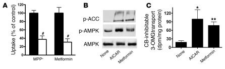
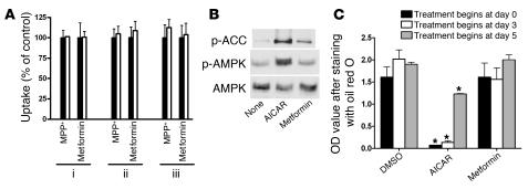
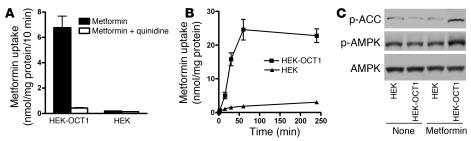
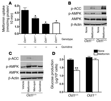
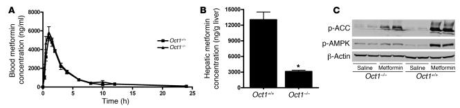
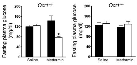
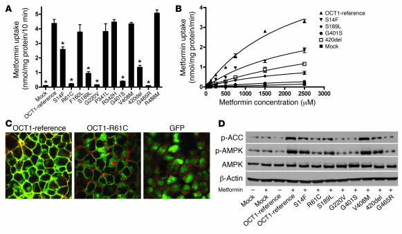
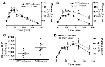
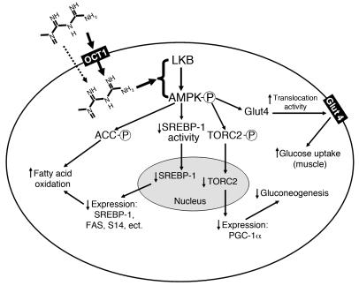
Metformin is among the most widely prescribed drugs for the treatment of type 2 diabetes. Organic cation transporter 1 (OCT1) plays a role in the hepatic uptake of metformin, but its role in the therapeutic effects of the drug, which involve activation of AMP-activated protein kinase (AMPK), is unknown. Recent studies have shown that human OCT1 is highly polymorphic. We investigated whether OCT1 plays a role in the action of metformin and whether individuals with OCT1 polymorphisms have reduced response to the drug. In mouse hepatocytes, deletion of Oct1 resulted in a reduction in the effects of metformin on AMPK phosphorylation and gluconeogenesis. In Oct1-deficient mice the glucose-lowering effects of metformin were completely abolished. Seven nonsynonymous polymorphisms of OCT1 that exhibited reduced uptake of metformin were identified. Notably, OCT1-420del (allele frequency of about 20% in white Americans), previously shown to have normal activity for model substrates, had reduced activity for metformin. In clinical studies, the effects of metformin in glucose tolerance tests were significantly lower in individuals carrying reduced function polymorphisms of OCT1. Collectively, the data indicate that OCT1 is important for metformin therapeutic action and that genetic variation in OCT1 may contribute to variation in response to the drug.


Figures

Figure 1
Figure 1. OCT activity and metformin responses in Clone 9 cells.
(A) Clone 9 cells exhibit OCT activity, as demonstrated by significantly reduced uptake of the typical OCT substrate MPP+ (1 μM) and metformin (250 μM) in the presence of the OCT inhibitor quinidine (100 μM; white bars) versus those without quinidine (control; black bars). The uptake times were 2 minutes for MPP+ and 10 minutes for metformin. The percentage uptake of the control was used to normalize the results for the 2 compounds in the same figure. #P < 0.001 versus respective controls (2-tailed Student’s t test). (B) Metformin (2 mM) stimulated the phosphorylation of AMPK and ACC in Clone 9 cells. The AMPK activator AICAR (2 mM) was used as a positive control. Cell extracts were detected with polyclonal antibodies against phospho-ACC (Ser79), phospho-AMPKα (Thr172), and AMPKα. (C) Metformin increased 3-OMG transport in Clone 9 cells. The cells were treated with metformin (2 mM) or AICAR (2 mM; positive control) for 2 hours before initiation of 3-OMG transport. 3-OMG transport was measured as described in Methods. CB, cytochalasin B. *P < 0.01, **P < 0.05 versus no treatment (ANOVA and Dunnett’s procedure).
Figure 2
Figure 2. No OCT activity and metformin responses in 3T3-L1 cells.
(A) No OCT activity was detected in 3T3-L1 cells of various differential stages. Uptake studies were done as described for Figure 1A. 3T3-L1 cells were differentiated as described in Methods. The cells for uptake experiments were: (i) preadipocytes; (ii) 3T3-L1 cells differentiated for 5 days; and (iii) 3T3-L1 cells differentiated for 10 days. (B) Metformin (2 mM) had little effect on the phosphorylation of AMPK and ACC in 3T3-L1 cells. The AMPK activator AICAR (2 mM) was used as a positive control. Cell extracts were detected with polyclonal antibodies against phospho-ACC (Ser79), phospho-AMPKα (Thr172), and AMPKα. (C) Metformin (2 mM) treatment during differentiation did not affect lipid accumulation in 3T3-L1 cells, in contrast to the significant effects of AICAR treatment (2 mM). The cellular lipids were stained with oil red O, and lipid content was determined by measuring the OD of the dye extracted with isopropyl alcohol. *P < 0.01 versus 0.05% DMSO (2-tailed Student’s t test).
Figure 3
Figure 3. Overexpressing human OCT1 in HEK293 cells increases metformin uptake and metformin-stimulated AMPK phosphorylation.
(A) The uptake of metformin in HEK293 cells was markedly increased by stable overexpression of human OCT1 in the cells. The uptake experiments were performed as described in Methods and in Figure 1A. “HEK” represents empty vector–transfected cells. (B) The uptake of metformin in the HEK293 cells overexpressing human OCT1 was time dependent. (C) The phosphorylation of AMPK and ACC by metformin in HEK293 cells was markedly increased by stably overexpressing human OCT1 in the cells. The cells were treated with metformin (250 μM) for 1 hour. Cell extracts were detected with polyclonal antibodies against phospho-ACC (Ser79), phospho-AMPKα (Thr172), and AMPKα.
Figure 4
Figure 4. Oct1 deletion results in reduced metformin uptake and response in primary hepatocytes from mice.
(A) Metformin uptake was lower in the primary hepatocytes isolated from Oct1-knockout (Oct1–/–) mice than in those with a normal Oct1 allele (Oct1+/+ and Oct1+/–). The uptake of metformin (250 μM) was performed for 10 minutes in the presence or absence of 100 μM quinidine, where indicated. *P < 0.01 versus Oct1+/– without quinidine (ANOVA and Dunnett’s procedure). (B) Metformin resulted in less phosphorylation of AMPK and ACC in Oct1–/– hepatocytes than in Oct1+/+ hepatocytes. The cellular extracts from primary hepatocytes treated with or without metformin (250 μM) for 4.5 hours were detected with polyclonal antibodies against phospho-ACC (Ser 79), phospho-AMPKα (Thr172), AMPKα, and β-actin. (C) Treatment with the OCT inhibitor quinidine reduced the stimulation of AMPK phosphorylation and thus ACC phosphorylation by metformin in Oct1+/+ hepatocytes. Where indicated, 100 μM quinidine was added 30 minutes before metformin (250 μM) treatment (Met + quin). (D) Metformin suppressed glucagon-stimulated glucose production in Oct1+/+ hepatocytes, with no effect in Oct1–/– hepatocytes. Metformin (1 mM) was added 2 hours before glucose measurement. The primary hepatocytes were isolated and cultured as described in Methods. **P < 0.001 versus no treatment (2-tailed Student’s test).
Figure 5
Figure 5. Oct1 deletion results in reduced hepatic metformin accumulation and phosphorylation of AMPK and ACC in mice receiving oral doses of metformin.
(A) The pharmacokinetics of metformin was similar in age-matched Oct1+/+ mice and Oct1–/– mice after an oral dose. Shown here are blood metformin concentration–time profiles. The mice (n = 4 per group) were given an oral dose of metformin (15 mg/kg containing 0.2 mCi/kg of [14C]metformin), approximating the single dose of 1,000 mg in humans. The radioactivity in blood was determined and converted to mass amounts. Data represent mean ± SD. (B) Hepatic metformin accumulation after an oral dose was much higher for Oct1+/+ mice than for age-matched Oct1–/– mice. The mice (n = 4 per group) were sacrificed 1 hour after the oral dose, and the livers were removed immediately. The radioactivity determined in liver homogenates was converted to mass amounts. Data represent mean ± SD. *P < 0.001 versus Oct1+/+ (2-tailed Student’s t test). (C) OCT1 was required for metformin to fully stimulate hepatic AMPK phosphorylation and ACC phosphorylation in mice. A daily dose of metformin (50 mg/kg) or saline was administered i.p. for 3 consecutive days to 10-week-old male mice. The mice were sacrificed 1 hour after the i.p. administration on the third day. Liver extracts were detected with polyclonal antibodies against phospho-ACC (Ser79), phospho-AMPKα (Thr172), and β-actin.
Figure 6
Figure 6. OCT1 is required for metformin to lower fasting plasma glucose levels in mice.
The 6-week-old Oct1+/+ mice and Oct1–/– mice (n = 5–8 per group) were administered a high-fat diet for 8 weeks, and 18-hour fasting plasma glucose concentrations were measured before (black bars; day 0) and after (white bars) 5 days of i.p. treatment with saline or metformin (50 mg/kg each day). Data represent mean ± SD. *P = 0.012 versus day 0 (2-tailed Student’s t test).
Figure 7
Figure 7. OCT1 genetic variants are associated with different accumulation rates and responses to metformin in stably transfected HEK293 cells.
(A) Uptake of [14C]metformin by cell lines stably expressing human OCT1 and its variants. Cells expressing OCT1 and its variants were incubated with [14C]metformin (250 μM) for 10 minutes. Seven OCT1 variants exhibited reduced metformin uptake as compared with OCT1-reference. Data are expressed as mean ± SD for samples analyzed in quadruplicate. *P < 0.001 compared with the reference (2-tailed Student’s t test). (B) Metformin kinetics in cell lines expressing reduced function variants of OCT1. Four of the reduced function variants shown in A had enough activity to allow us to perform kinetic studies with metformin. The metformin uptake data at 8 different concentrations are plotted. The variants had significantly different Vmax values, with a similar Michaelis-Menten constant (Km) (Table 1). (C) OCT1-R61C tagged with GFP exhibits reduced membrane and enhanced cytoplasmic localization. GFP fusion constructs were generated for OCT1-reference and OCT1-R61C, which is common in human populations (13), and used to generate stable cell lines using Flp-In-293 cells. The plasma membrane was stained using Alexa Fluor 594 conjugated to wheat germ agglutinin, and cells were visualized by confocal microscopy. Original magnification, ×100. (D) Metformin-stimulated AMPK phosphorylation and ACC phosphorylation in cell lines stably overexpressing human OCT1 and its variants. The cells were treated with metformin (1 mM) for 1 hour, washed with blank medium, and then incubated for 5 hours before harvest. Immunoblots were performed against phospho-ACC (Ser79), phospho-AMPKα (Thr172), AMPKα, and β-actin.
Figure 8
Figure 8. OCT1 genetic variants are associated with different responses to metformin in healthy human volunteers.
(A) The time course of plasma glucose concentrations for a baseline OGTT without metformin treatment in healthy subjects having only reference OCT1 alleles (n = 8) and those having at least 1 reduced-function OCT1 allele (n = 12). The data are expressed as mean ± SEM. (B) The time course of plasma glucose concentrations for OGTT after metformin treatment in the same healthy subjects represented in A. The data are expressed as mean ± SEM; *P < 0.05 compared with volunteers with only reference OCT1 alleles (unpaired Student’s t test). (C) The glucose exposure with OGTT (AUC) after metformin treatment for healthy subjects represented in B. The horizontal lines represent mean values for the 2 groups. The mean value for volunteers with only reference OCT1 alleles is significantly lower than that for the variant group. P = 0.004 (unpaired Student’s t test). (D) The time course of insulin levels during the OGTT after metformin administration in the same healthy individuals represented in A. The data are expressed as mean ± SEM; *P < 0.05 compared with individuals with only OCT1-reference alleles (unpaired 1-tailed Student’s t test).
Figure 9
Figure 9. Mechanism of metformin action in cells.
By controlling the intracellular concentrations, OCT1 is a direct determinant of metformin pharmacological effects in the liver (bold arrow). Passive diffusion and other transporters may account for small portion of hepatic uptake of metformin (dashed arrow). Other transporters may control metformin uptake into other tissues, such as skeletal muscle. Factors such as genetic variation in transporter genes may alter transporter activity and thus metformin response. LKB, alias of serine-threonine kinase 11 (STK11); PGC-1α, peroxisome proliferator activated receptor γ coactivator 1 α; TORC2, target of rapamycin complex 2.

Comment in

References

    1. Kirpichnikov D., McFarlane S.I., Sowers J.R. Metformin: an update. Ann. Intern. Med. 2002;137:25–33. - PubMed
    1. Lin H.Z., et al. Metformin reverses fatty liver disease in obese, leptin-deficient mice. Nat. Med. 2000;6:998–1003. - PubMed
    1. Baillargeon J.P., Iuorno M.J., Nestler J.E. Insulin sensitizers for polycystic ovary syndrome. Clin. Obstet. Gynecol. 2003;46:325–340. - PubMed
    1. Zhou G., et al. Role of AMP-activated protein kinase in mechanism of metformin action. J. Clin. Invest. 2001;108:1167–1174. doi: 10.1172/JCI200113505. - DOI - PMC - PubMed
    1. Abbud W., et al. Stimulation of AMP-activated protein kinase (AMPK) is associated with enhancement of Glut1-mediated glucose transport. Arch. Biochem. Biophys. 2000;380:347–352. - PubMed

Publication types