. 2007 May 14:(18):1798-800.
doi: 10.1039/b703195e. Epub 2007 Mar 28.

Human frataxin: iron and ferrochelatase binding surface

Affiliations

Human frataxin: iron and ferrochelatase binding surface

Krisztina Z Bencze et al. Chem Commun (Camb). .

Abstract

The coordinated iron structure and ferrochelatase binding surface of human frataxin have been characterized to provide insight into the protein's ability to serve as the iron chaperone during heme biosynthesis.


Figures
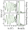
Fig. 1
Fig. 1
EXAFS of frataxin bound iron. Raw EXAFS (black) and simulations (green) of 1 (A) and 2 (C) Fe bound. Fourier transforms of raw (black) and simulated (green) EXAFS of 1 (B) and 2 (D) Fe bound.
Fig. 2
Fig. 2
Human frataxin residues perturbed at their amide position upon forming a complex with iron and ferrochelatase. Unshifted (green) and shifted (red) residues identified on the apo-protein’s structure. Yellow residues are line broadened beyond recognition resulting from Fe binding.
Fig. 3
Fig. 3
Docking simulation of frataxin (Ftx) (green) with the ferrochelatase (Frch) dimer. Frataxin interacts with ferrochelatase on the matrix exposed side of the protein dimer during iron delivery (yellow ball). Delivered iron would then be available to be transported down ferrochelatase’s substrate channel (red arrow) towards the enzyme’s active site (yellow oval) located on the membrane side of the molecule.
Fig. 4
Fig. 4
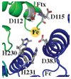
Expanded view of metal binding residues at the proposed frataxin (green)/ferrochelatase molecule 2 (blue) interface.

References

    1. Delatycki MB, Williamson R, Forrest SM. J. Med. Genet. 2000;37:1–8. - PMC - PubMed
    1. Bencze KZ, Kondapalli KC, Cook JD, McMahon S, Millan-Pacheco C, Pastor N, Stemmler TL. Crit. Rev. Biochem. Mol. Biol. 2006;41:269–291. - PMC - PubMed
    1. Adamec J, Rusnak F, Owen WG, Naylor S, Benson LM, Gacy AM, Isaya G. Am. J. Hum. Genet. 2000;67:549–562. - PMC - PubMed
    1. Nair M, Adinolfi S, Pastore C, Kelly G, Temussi P, Pastore A. Structure (Cambridge, MA, U. S.) 2004;12:2037–2048. - PubMed
    1. He Y, Alam SL, Proteasa SV, Zhang Y, Lesuisse E, Dancis A, Stemmler TL. Biochemistry. 2004;43:16254–16262. - PMC - PubMed

Publication types