. 2007;35(10):3453-64.
doi: 10.1093/nar/gkm239. Epub 2007 May 3.

Transcription of the human cell cycle regulated BUB1B gene requires hStaf/ZNF143

Affiliations

Transcription of the human cell cycle regulated BUB1B gene requires hStaf/ZNF143

Evelyne Myslinski et al. Nucleic Acids Res. 2007.

Abstract

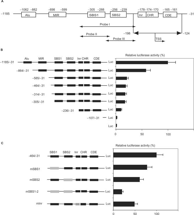
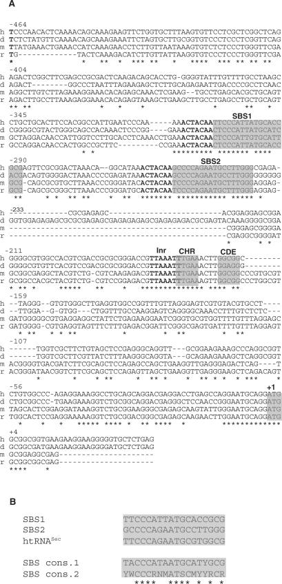
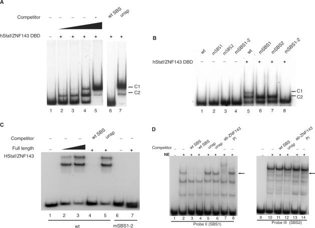
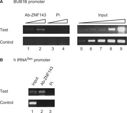
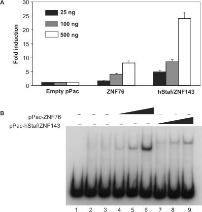
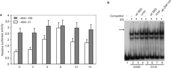
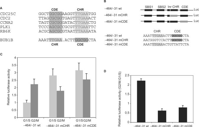
BubR1 is a key protein mediating spindle checkpoint activation. Loss of this checkpoint control results in chromosomal instability and aneuploidy. The transcriptional regulation of the cell cycle regulated human BUB1B gene, which encodes BubR1, was investigated in this report. A minimal BUB1B gene promoter containing 464 bp upstream from the translation initiation codon was sufficient for cell cycle regulated promoter activity. A pivotal role for transcription factor hStaf/ZNF143 in the expression of the BUB1B gene was demonstrated through gel retardation assays, transient expression of mutant BUB1B promoter-reporter gene constructs and chromatin immunoprecipitation assay. Two phylogenetically conserved hStaf/ZNF143-binding sites (SBS) were identified which are indispensable for BUB1B promoter activity. In addition, we found that the domain covering the transcription start sites contains conserved boxes homologous to initiator (Inr), cell cycle dependent (CDE) and cell cycle genes homology regions (CHR) elements. Mutations within the CDE and CHR elements led to diminished cell cycle regulation of BUB1B transcription. These results demonstrate that BUB1B gene transcription is positively regulated by hStaf/ZNF143, a ubiquitously expressed factor, and that the CDE-CHR tandem element was essential for G2/M-specific transcription of the BUB1B gene.


Figures

Figure 1.
Figure 1.
Identification of the human BUB1B gene promoter and mutation analysis of promoter elements. (A) Schematic representation of the region located 5′ to the BUB1B gene. The different elements identified are boxed. Numbering is relative to the translation initiation codon. The double-headed arrow depicts the region containing the various TSS. Gel shift probes I, II and III are diagrammed below the promoter. (B) Schematic representation of the 5′-deleted BUB1B promoter-luciferase constructs and their activities in COS-7 cells. Cells were transiently transfected with the 5′-deleted BUB1B promoter-luciferase (Luc) constructs and assayed for luciferase activity. Values are given as relative luciferase activity normalized to 100 for the longest promoter construct −1185/−31. Data are presented as the mean +/−SD of three separate experiments. (C) Schematic representation of BUB1B promoter-luciferase constructs mutated in the Inr-like motif, SBS1 and SBS2 elements and their activities in COS-7 transfected cells. Wild-type and mutant elements are boxed in black and gray, respectively. The relative luciferase activity in % was normalized to 100 with the −464/−31 construct. Data are presented as in (B).
Figure 2.
Figure 2.
Cross-species conservation of putative promoter elements in the promoter regions of BUB1B homologous genes. (A) Nucleotide sequence comparison of the human (h), dog (d), mouse (m) and rat (r) BUB1B promoters. Multiple sequence alignments performed with Clustal W covering the −464/+35 part of the human promoter. Identical nucleotides are indicated with a star. Numbering is relative to the translation initiation codon. The SBS1, SBS2, elements homologous to CHR and CDE motifs, and translational start codon are highlighted in gray. The Inr-like motif and ACTACAA submotif are in bold. (B) Comparison of the SBS1 and SBS2 sequences of the BUB1B promoter, SBS of the human tRNASec (htRNASec) gene (11) and SBS consensus sequences determined by binding site selection at highly (Staf cons.1) (35) or moderately stringent selection conditions (Staf cons.2) (11), (Y, W, R, N, M and S stand for T/C, A/T, A/G, any nucleotide, A/C and G/C, respectively). Identical nucleotides are indicated with a star.
Figure 3.
Figure 3.
hStaf/ZNF143-binding assays on wild-type and mutant versions of the BUB1B promoter. (A) Gel retardation assay with a fragment encompassing positions −342/−195 of the BUB1B promoter. The 32P labeled DNA (probe I) was incubated in the absence (lane 1) or presence of increasing amounts of hStaf/ZNF143 DBD (lanes 2–5). The reactions in lanes 6 and 7 were performed with the same amount of protein as in lane 5 but in the presence of a 1000-fold excess of unlabeled specific (wt SBS) and unspecific competitors (unsp). Binding assays in lanes 1–5 and 6, 7 were performed in separate experiments. C1 and C2: complexes containing one and two proteins, respectively. (B) A 148-bp 5′ end-labeled fragment containing the wild-type or mutant versions of the SBS was used in the binding studies. Lanes 1–4, no protein added. Lanes 5–8 binding assays with the same amount of DBD. Probes are indicated above the lanes. (C) The wt (lanes 1–5) or the SBS1 and SBS2 mutant version borne by probe I (lanes 6 and 7) were incubated in the absence (lanes 1 and 6) or presence of increasing amounts of rabbit reticulocyte lysate containing full-length hStaf/ZNF143 (lanes 2 and 3). Lane 7 contained the same amount of protein as in lane 3. Lanes 4 and 5 were performed with the same amount of protein as in lane 3 but in the presence of a 1000-fold excess of unlabeled specific (lane 4, wt SBS) and unspecific competitor (lane 5, unsp), respectively. C1 corresponds to both sites of the probe saturated with the protein; the protein binds to one of the sites only in C2. (D) Gel retardation assay with the wt probes II (lanes 1–8) or III (lanes 9–14) in the absence (lanes 1 and 9) or presence (lanes 2–8 and 10–14) of HeLa cell nuclear extracts (NE). The reactions in lanes 3, 4 and 11, 5, 6 and 12, 7 and 13, 8 and 14 were performed in the presence of unlabeled specific competitor (wt SBS), unspecific competitor (unsp), anti-hStaf/ZNF143 and pre-immune antibody, respectively. The specific competitor was added at a 500-fold (lane 3) and 1000-fold molar excess (lanes 4, 11). A 500 and 1000-fold molar excess of unspecific competitor was contained in lanes 5 and 6, 12 respectively. The arrow points to the complexes mentioned in the text.
Figure 4.
Figure 4.
hStaf/ZNF143 is associated to the BUB1B gene promoter in HeLa cells. Genomic DNA fragments, recovered from input material or immunoprecipitated with hStaf/ZNF143 or pre-immune antibodies, were subjected to PCR amplification with primer pairs specific for the promoter or upstream regions. (A) PCR analysis of the BUB1B promoter. Lanes 1, 2 and 3, 4: two different amounts (1 and 5%) of immunoprecipitated DNA with hStaf/ZNF143 (Ab-ZNF143) or pre-immune antibodies (Pi), respectively. Lanes 5–9: increasing amounts of input material (0.002, 0.01, 0.05, 0.2 and 1%) were analyzed to demonstrate that the assays were within the linear range of PCR amplification. PCR primer pairs were specific for the promoter region of the BUB1B gene (test assay) or for a unique region located ∼2.5 kbp upstream of the BUB1B gene (control assay). (B) PCR amplification was performed with a primer pair specific for the human tRNASec (htRNASec) gene promoter (test assay) or a region located 2.4 kbp upstream of it (control assay). Lanes 1, PCR performed with 0.01% of input DNA. Lanes 2 and 3, PCR performed with 1% of DNA recovered from ChIP.
Figure 5.
Figure 5.
hStaf/ZNF143 and ZNF76 transactivate the human BUB1B promoter in Drosophila Schneider cells. (A) SL2 cells were cotransfected with the −464/−31 luciferase reporter gene construct and increasing amounts of empty pPac, pPac-hStaf/ZNF143 or pPac-ZNF76 expression vectors (25 ng, solid boxes; 100 ng, gray boxes; 500 ng, open boxes). Data are expressed as the fold of induction of luciferase activity normalized to that obtained following co-transfection of the reporter with the empty vector pPac. Values are the mean +/−SD of at least three independent transfection experiments. (B) Expression of ZNF76 and hStaf/ZNF143 in transfected SL2 cells. Expression of the proteins in SL2 cells was confirmed by gel retardation assays. The wt probe II containing SBS1 was incubated with untransfected SL2 extracts (lane 2), transfected SL2 extracts with empty pPac (lane 3), transfected SL2 extracts with pPac-ZNF76 (lanes 4–6) or pPac-hStaf/ZNF143 (lanes 7–9). Lanes 4 and 7, 5 and 8, 6 and 9: probes incubated with extracts from SL2 cells transfected with 25, 100 and 500 ng of effectors, respectively.
Figure 6.
Figure 6.
The luciferase activity arising from the construct containing the BUB1B promoter is regulated during the cell cycle. (A) COS-7 cells were transiently transfected with −464/−31 (open boxes) or −464/−196 (solid boxes) constructs. Cells were synchronized with a double thymidine block and released (time 0) and harvested at the indicated times for luciferase assays. Data are presented as the mean +/−SD of three independent experiments. (B) Gel retardation assay with the wt probe II in the absence (lane 1) or presence of COS-7 nuclear extracts from cells synchronized in G2/M (lanes 2–4) or G1/S (lanes 5–8). Reactions in lanes 3 and 6, 4 and 7, 8 were performed in the presence of unlabeled specific competitor (wt SBS), unspecific competitor (unsp) and anti-hStaf/ZNF143 antibody, respectively. Position of the specific complex is indicated with an arrow.
Figure 7.
Figure 7.
Effects of mutations of the CDE and CHR elements on the BUB1B promoter activity. (A) Alignment of the CDC25C, CDC2, CCNA2, PLK1 and RB6K promoter sequences in the region of the CDE and CHR elements (25–27). CDE and CHR elements are highlighted in gray. The bottom line displays the part of the BUB1B promoter sequence harboring high identity with the CDE and CHR motifs. (B) Schematic representation of the BUB1B luciferase constructs and sequences of the wild-type and mutant promoters. Here, −464/−31 mCDE and −464/−31 mCHR correspond to substitutions including the conserved GGCGG and TTGAA, respectively. (C) Transient transfection experiments into COS-7 cells with the wild-type −464/−31 and mutant −464/−31 mCHR and −464/−31 mCDE constructs. Following transfection, cells were treated with thymidine to block exit from G1/S (open boxes) or with nocodazole to block that from G2/M (solid boxes). Cells were harvested and assessed for luciferase activity. (D) The relative luciferase activity of cells in G2/M versus G1/S is given for the wild-type and mutant constructs. Data are presented as the mean +/−SD of three independent experiments.

Similar articles

Cited by

References

    1. Lengauer C. Cancer. An unstable liaison. Science. 2003;300:442–443. - PubMed
    1. Bharadwaj R, Yu H. The spindle checkpoint, aneuploidy, and cancer. Oncogene. 2004;23:2016–2027. - PubMed
    1. Lengauer C, Kinzler KW, Vogelstein B. Genetic instabilities in human cancers. Nature. 1998;396:643–649. - PubMed
    1. Dai W, Wang Q, Liu T, Swamy M, Fang Y, Xie S, Mahmood R, Yang YM, Xu M, et al. Slippage of mitotic arrest and enhanced tumor development in mice with BubR1 haploinsufficiency. Cancer Res. 2004;64:440–445. - PubMed
    1. Rao CV, Yang YM, Swamy MV, Liu T, Fang Y, Mahmood R, Jhanwar-Uniyal M, Dai W. Colonic tumorigenesis in BubR1+/−ApcMin/+ compound mutant mice is linked to premature separation of sister chromatids and enhanced genomic instability. Proc. Natl Acad. Sci. USA. 2005;102:4365–4370. - PMC - PubMed

Publication types

MeSH terms