. 2007;35(10):3407-19.
doi: 10.1093/nar/gkm206. Epub 2007 May 3.

Sequences in attB that affect the ability of phiC31 integrase to synapse and to activate DNA cleavage

Affiliations

Sequences in attB that affect the ability of phiC31 integrase to synapse and to activate DNA cleavage

Milind Gupta et al. Nucleic Acids Res. 2007.

Abstract

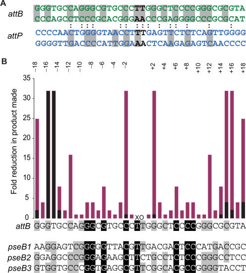
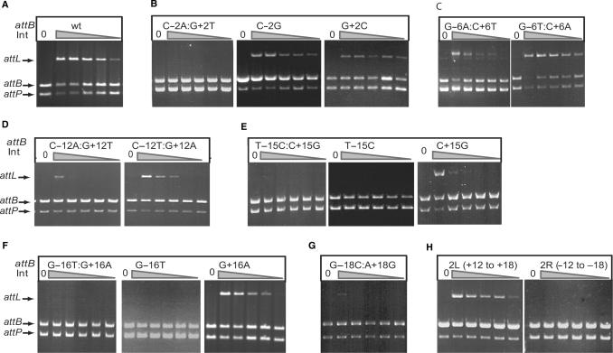
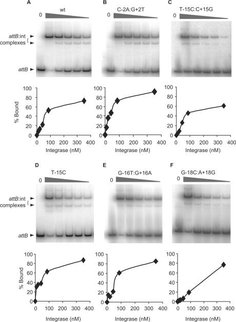
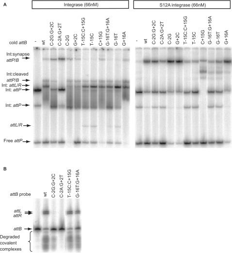
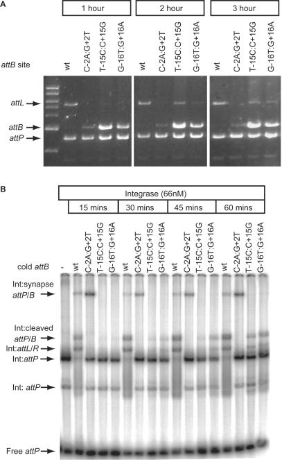
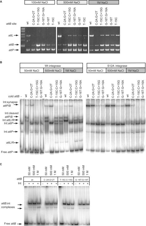
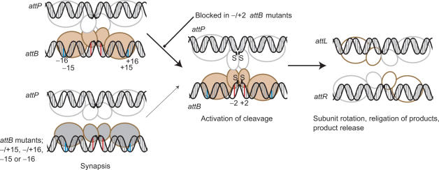
Phage integrases are required for recombination of the phage genome with the host chromosome either to establish or exit from the lysogenic state. C31 integrase is a member of the serine recombinase family of site-specific recombinases. In the absence of any accessory factors integrase is unidirectional, catalysing the integration reaction between the phage and host attachment sites, attP x attB to generate the hybrid sites, attL and attR. The basis for this directionality is due to selective synapsis of attP and attB sites. Here we show that mutations in attB can block the integration reaction at different stages. Mutations at positions distal to the crossover site inhibit recombination by destabilizing the synapse with attP without significantly affecting DNA-binding affinity. These data are consistent with the proposal that integrase adopts a specific conformation on binding to attB that permits synapsis with attP. Other attB mutants with changes close to the crossover site are able to form a stable synapse but cleavage of the substrates is prevented. These mutants indicate that there is a post-synaptic DNA recognition event that results in activation of DNA cleavage.


Figures

Figure 1.
Figure 1.
ϕC31 attB and attP sites. (A) The double-stranded DNA sequences of the S. coelicolor attB site (green) and the attP site (blue) are shown. The crossover dinucleotides are shown in black. The colons connecting the two sequences indicate the positions of sequence identity between the aligned attB and attP sites. The grey shading indicates the positions where sequence conservation can be detected between the attB or attP sites and their pseudo-sites from Streptomyces or Mycobacteria (pseudo-attB sites) or from human or mouse cell lines (pseudo-attP sites) (9,41–43). (B) Summary of mutation scanning in attB. The attB site is shown as a single-strand sequence where each base acts as point on the x-axis of a histogram. The y-axis shows the fold reduction in product made when mutations are introduced in attB. The positions are annotated according to the numbering shown. The activities of attB sites with double mutations at symmetrical positions (eg −/+1, −/+2, etc.) are shown in pink and the activities of mutants with single mutations are shown in black. The data for the summary graph were calculated from the estimated absolute activities shown in Table 1, Figure 2 and Supplementary Data, Figure S1. Beneath the attB sequence, three of the S. coelicolor pseudo-attB sites [pseB1, pseB2 and pseB3 (41)] are shown for comparison with the wild-type attB. The four sites have been aligned and are shaded according to whether there is 100% identity (black background and white text) or 75% identity (grey background) between the sites.
Figure 2.
Figure 2.
Recombination activities of attB mutant sites. Recombination activities are shown for the wild-type attB site (A), mutant sites at position 2 (B), 6 (C), 12 (D), 15 (E), 16 (F) 18 (G). Panel H shows the activities of partially symmetrized attB sites that contain the right sequence between +12 and +18 changed to the same sequence as on the left (−12 to −18), 2L (+12 to +18) or vice versa, 2R (−12 to −18). Recombination assays were performed using the standard plasmid assay containing the plasmid indicated in each panel and pRT702 encoding attP. The concentrations of integrase used for each set of six reactions in panels A to C and E, F and H was 0, 441, 110, 55, 27 and 14 nM. The concentrations of integrase used for each set of six reactions in panels D and G was 0, 351, 87, 43, 21 and 10 nM.
Figure 3.
Figure 3.
Binding affinities by integrase for the wild-type and mutant attB sites. Integrase was incubated with radiolabelled wild type (panel A) and mutant attB sites (panels B–F). In each panel, the phosphorimage shows the complexes obtained with increasing integrase concentrations and, below, the quantitative analysis of the% bound versus the concentration of integrase. Only the −/+15 mutant (T-15C:C+15G) and the −/+18 mutant (G-18C:A+18G) sites showed reduced binding affinities for integrase under the conditions used. A summary of the integrase concentrations required for 50% binding of the different attB mutants is shown in Table 2.
Figure 4.
Figure 4.
Synapse assays with wild-type and mutant attB sites. Panel (A) Radiolabelled attP was incubated with wild-type (left panel) or the catalytically inactive integrase, S12A, (right panel) and a cold partner fragment containing wild-type attB (wt) or the indicated mutant attB sites. The arrows show the positions of the synapse containing the radiolabelled substrate, the cold partner fragment and integrase (Int:synapse attP/B), the covalently linked cleaved substrate (Int:cleaved attP/B), the shifted and free products (Int:attL/R and attL/R, respectively) and the positions of the attP bound only to integrase (two complexes labelled Int:attP) or free (attP). Panel (B) The protease subtilisin was used to reveal the extent of cleavage of attB sites and the products formed during the synapse assay. Arrows show the positions of the products, attL and attR, the radiolabelled substrate and attB. The smear of radioactivity migrating faster than the free probe results from subtilisin treated cleaved covalently linked complexes.
Figure 5.
Figure 5.
The rate of recombination with mutant attB sites T-15C:C+15G and G-16T:G+16A is greatly reduced. Panel (A) shows the appearance of products from recombination assays using T-15C:C+15G and G-16T:G+16A as substrates after prolonged incubation. Plasmids encoding the wild-type attB (wt) or the indicated attB mutants were incubated with pRT702 (attP) for 1, 2 or 3 h at 30°C and then the products analysed by restriction and agarose gel electrophoresis. After 2 and 3 h some product (attL) is visible in the lanes containing the −/+15 and −/+16 mutations. Panel (B) shows the time-dependent appearance of recombination intermediates when wild-type attB (wt) was used compared to C-2A:G+2T, T-15C:C+15G or G-16T:G+16A. The −/+2 mutant site rapidly forms a synapse (Int:synapse attP/B) and thereafter the reaction is blocked. The −/+15 and −/+16 attB sites slowly accumulated the cleaved intermediate (Int:cleaved attP/B) and some shifted product complexes (Int:attL/R). The remaining complexes on the gel are as described in Figure 4.
Figure 6.
Figure 6.
High NaCl concentrations can partially suppress the recombination defective phenoytpe of the mutations. Panel (A) shows the appearance of products from recombination assays using T-15C:C+15G, G-16T:G+16A, G-16T and T-15C as substrates after incubation in either 500 mM or 1 M NaCl. Plasmids encoding the wild-type attB (wt), or the above mutants were incubated with pRT702 (attP) in recombination buffer adjusted to 100 mM, 500 mM or 1 M NaCl. After digesting with HindIII the DNA was separated in an agarose gel. The appearance of the 5435 bp fragment encoding attL is indicative of recombination. Panel (B) shows the synapse assays using the wild-type attB (wt), C-2A:G+2T, T-15C:C+15G and G-16T:G+16A under different NaCl conditions with wild type (left panel) or S12A integrase (right panel) with labelled attP. The complexes are annotated as described in Figure 4. −/+15 and −/+16 mutant attB sites accumulated both the cleaved complex (Int:cleaved attP/B), the shifted products (Int:attL/R) and released some free product (attL/R) with 500 mM and 1 M NaCl with the wild-type integrase. The synapse however as indicated using the S12A integrase did not become more abundant in high NaCl buffer, if anything it reduced. Panel (C) shows that the binding affinity of attB sites for integrase in the presence of different NaCl concentrations. Radiolabelled attB sites were incubated in binding buffer containing 50 mM, 500 mM or 1 M NaCl. Integrase was added at 66 nM. The shifted attB complexes are indicated by arrows. Only at 1 M NaCl, there was a slight increase in the free DNA for all four attB sites.
Figure 7.
Figure 7.
Model for the mechanism of integrase. The integrase subunits are shown with a small N-terminal (catalytic) domain through which the subunits may dimerize (26) and a large C-terminal domain that we propose recognizes the sequences in the outer flanks of the recombination sites. These recognition events, specifically −15 and −16 (annotated as blue bars) in attB, lead to an ‘induced fit’ or stabilization of a specific conformation of integrase that enables synapsis with integrase bound to attP. Different conformations of integrase bound to either attB or attP are shown as different colours. The synaptic interface via the N-terminal catalytic domains is indicated based on the resolvase precedent; there is no evidence to indicate that the C-terminal domains could also participate in a synaptic interface. Mutations at positions −15 or −16 (such as in T-15C:C+15G, G-16T:G+16A), T-15C or G-16T do not induce the conformation of integrase that can form a stable synapse with attP so the rate of reaction decreases (thin arrow). Mutations at −/+2 in attB (red bars) are severely inhibited in cleavage but are capable of forming a stable synapse. These mutants indicate that the formation of the synaptic complex is followed by a well-defined activation step that results in concerted DNA cleavage. After strand exchange integrase is bound to the hybrid sites and adopts a conformation that cannot synapse attL and attR. The putative tetrameric complex rapidly dissociates to binary complexes containing integrase and either attL or attR. See text for more details.

Similar articles

Cited by

References

    1. Bateman JR, Lee AM, Wu CT. Site-specific transformation of Drosophila via ϕC31 integrase-mediated cassette exchange. Genetics. 2006;173:769–777. - PMC - PubMed
    1. Andreas S, Schwenk F, Kuter-Luks B, Faust N, Kuhn R. Enhanced efficiency through nuclear localization signal fusion on phage ϕC31-integrase: activity comparison with Cre and FLPe recombinase in mammalian cells. Nucleic Acids Res. 2002;30:2299–2306. - PMC - PubMed
    1. Belteki G, Gertsenstein M, Ow DW, Nagy A. Site-specific cassette exchange and germline transmission with mouse ES cells expressing ϕC31 integrase. Nat Biotechnol. 2003;21:321–324. - PubMed
    1. Dafhnis-Calas F, Xu Z, Haines S, Malla SK, Smith MCM, Brown WR. Iterative in vivo assembly of large and complex transgenes by combining the activities of ϕC31 integrase and Cre recombinase. Nucleic Acids Res. 2005;33:e189. - PMC - PubMed
    1. Khan MS, Khalid AM, KMalik KA. Phage ϕC31 integrase: a new tool in plastid genome engineering. Trends in Plant Science. 2005;10:1–3. - PubMed

Publication types