. 2007 Aug 1;582(Pt 3):1337-48.
doi: 10.1113/jphysiol.2007.132639. Epub 2007 May 3.

The role of the actin cytoskeleton in oxytocin and vasopressin release from rat supraoptic nucleus neurons

Affiliations

The role of the actin cytoskeleton in oxytocin and vasopressin release from rat supraoptic nucleus neurons

Vicky A Tobin et al. J Physiol. .

Abstract

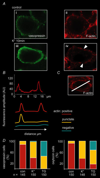
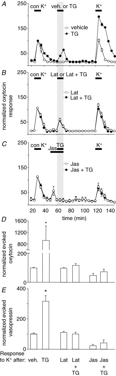
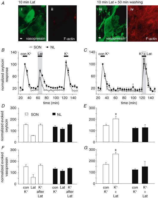
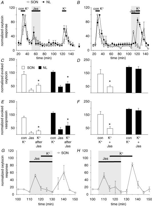
Magnocellular neurons of the supraoptic nucleus (SON) can differentially control peptide release from the somato/dendritic and axon terminal compartment. Dendritic release can be selectively regulated through activation of intracellular calcium stores by calcium mobilizers such as thapsigargin (TG), resulting in preparation (priming) of somato/dendritic peptide pools for subsequent activity-dependent release. As dynamic modulation of the actin cytoskeleton is implicated in secretion from synaptic terminals and from several types of neuroendocrine cells, we studied its involvement in oxytocin and vasopressin release from SON neurons. Confocal image analysis of the somata revealed that the normally continuous cortical band of F-actin is disrupted after high potassium (K(+), 50 mm) or TG (200 nm) stimulation. The functional importance of actin remodelling was studied using cell-permeable actin polymerizing (jasplakinolide, 2 microm) or depolymerizing agents (latrunculin B, 5 microm) to treat SON and neural lobe (NL) explants in vitro and measure high K(+)-induced oxytocin and vasopressin release. Latrunculin significantly enhanced, and jasplakinolide inhibited, high-K(+)-evoked somato/dendritic peptide release, while release from axon terminals was not altered, suggesting that high-K(+)-evoked release in the SON, but not the NL, requires depolymerization of the actin cytoskeleton. TG-induced priming of somato/dendritic release was also blocked by jasplakinolide and latrunculin, suggesting that priming involves changes in actin remodelling.


Figures

Figure 1
Figure 1. High potassium and thapsigargin induced reorganization of actin filaments in magnocellular neurons of the supraoptic nucleus (SON)
A, immunohistochemical detection of vasopressin and F-actin in control tissue (i and ii) and after treatment with high K+ (iii and iv). B, the trace shows the fluorescence signal profile for the line placed across the cell shown in C. Shown below are profiles of F-actin signals assigned as positive, punctate or negative. D, percentages of vasopressin and oxytocin neurons classified after high-K+ or TG treatment. Fluorescence amplitude is in arbitrary units (AU).
Figure 4
Figure 4. Effects of actin remodelling on thapsigargin-induced priming of oxytocin and vasopressin release in vitro
A, oxytocin release from isolated SONs evoked by depolarization with high K+ was strongly potentiated after thapsigargin-induced priming (TG, 0.2 μm). Latrunculin (B) or jasplakinolide (C) treatment did not induce priming and prevented the TG-induced priming in oxytocin (D) and vasopressin cells (E). Mean ±s.e.m., n = 6 per group, *P < 0.05 versus response to control high-K+ stimulus, one-way ANOVA.
Figure 2
Figure 2. Effects of actin depolymerization on oxytocin and vasopressin release from SON and neural lobe (NL) explants in vitro
A, treatment with latrunculin (Lat) removed the F-actin signal within the SON, which returned after a washout period. Actin depolymerization by latrunculin (5 μm) did not alter K+-induced release when given 50 min before (B), but significantly enhanced somato/dendritic release of oxytocin and vasopressin when given simultaneously with high-K+ (C). Evoked release of oxytocin (D and E) and vasopressin (F and G) for the two treatment procedures. Mean ±s.e.m., n = 6 per group, *P < 0.05 versus response to control high-K+ stimulus. One-way ANOVA was used for data shown in D and F and paired t test for data shown in E and G.
Figure 3
Figure 3. Effects of actin polymerization on oxytocin and vasopressin release from SON and NL explants in vitro
Actin polymerization by jasplakinolide (Jas; 2 μm) inhibited peptide release from the SON when given either 50 min before (A) or with high K+ (B). Oxytocin (C) and vasopressin (E) secretion from axon terminals of the NL was only reduced when jasplakinolide was given 50 min before high-K+ stimulation. Evoked release of oxytocin (C and D) and vasopressin (E and F) are shown for the two treatment procedures. G, to determine if inhibition of high-K+-induced secretion from SON explants during exposure to jasplakinolide was due to a depolarization-induced block (data from B); in H, high-K+ exposure was continued after termination of jasplakinolide treatment. Mean ±s.e.m., n = 6 per group, *P < 0.05 versus response to control high-K+ stimulus, paired t test.

Similar articles

Cited by

References

    1. Aunis D, Bader MF. The cytoskeleton as a barrier to exocytosis in secretory cells. J Exp Biol. 1988;139:253–266. - PubMed
    1. Bonder EM, Mooseker MS. Cytochalasin B slows but does not prevent monomer addition at the barbed end of the actin filament. J Cell Biol. 1986;102:282–288. - PMC - PubMed
    1. Borisy GG, Svitkina TM. Actin machinery: pushing the envelope. Curr Opin Cell Biol. 2000;12:104–112. - PubMed
    1. Bubb MR, Senderowicz AM, Sausville EA, Duncan KL, Korn ED. Jasplakinolide, a cytotoxic natural product, induces actin polymerization and competitively inhibits the binding of phalloidin to F-actin. J Biol Chem. 1994;269:14869–14871. - PubMed
    1. Cazalis M, Dayanithi G, Nordmann JJ. Hormone release from isolated nerve endings of the rat neurohypophysis. J Physiol. 1987;390:55–70. - PMC - PubMed

Publication types