Case Reports
. 2007 May;3(5):291-6.
doi: 10.1038/ncpneuro0493.

A patient with encephalitis associated with NMDA receptor antibodies

Affiliations
Case Reports

A patient with encephalitis associated with NMDA receptor antibodies

Lauren H Sansing et al. Nat Clin Pract Neurol. 2007 May.

Abstract

Background: A 34-year-old woman presented with headache, feverish sensation and anxiety, rapidly followed by homicidal ideation, aggressive agitation, seizures, hypoventilation, hyperthermia and prominent autonomic instability requiring intubation and sedation. She developed episodes of hypotension and bradycardia with periods of asystole lasting up to 15 seconds. Upon weaning off sedation, her eyes opened but she was unresponsive to stimuli. There was muscle rigidity, frequent facial grimacing, rhythmic abdominal contractions, kicking motions of the legs, and intermittent dystonic postures of the right arm.

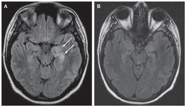
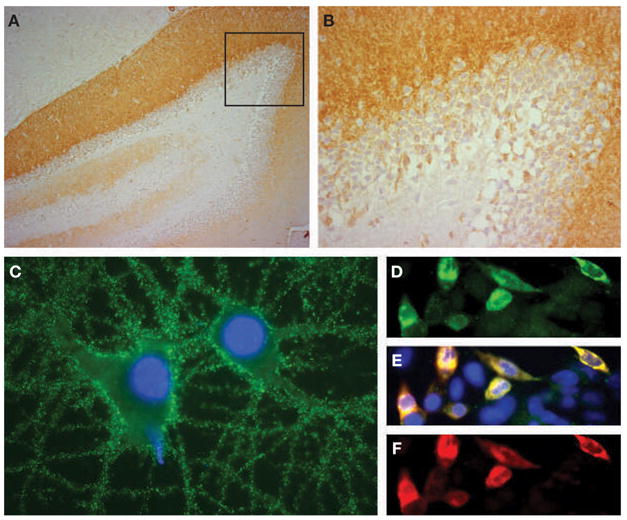
Investigations: Routine laboratory testing, toxicology screening, studies for autoimmune and infectious etiologies, brain MRI scan, lumbar puncture, electroencephalogram, whole-body CT scan, abdominal ultrasound, paraneoplastic and voltage-gated potassium channel antibody serologies, analysis of N-methyl-D-aspartate receptor antibodies.

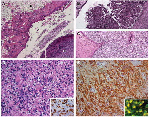
Diagnosis: Paraneoplastic encephalitis associated with immature teratoma of the ovary and N-methyl-D-aspartate receptor antibodies.

Management: Intensive care, mechanical ventilation, antiepileptics, laparotomy and left salpingo-oophorectomy, corticosteroids, plasma exchange, intravenous immunoglobulin, cyclophosphamide, physical therapy, and chemotherapy.


Figures

Figure 1
Figure 1
MRI scan of the patient at symptom presentation and follow-up. (A) MRI fluid-attenuated inversion recovery (FLAIR) obtained at symptom presentation demonstrates bilateral medial temporal lobe hyperintense signal, predominantly involving the left hippocampus (arrows). (B) Follow-up MRI obtained during recovery, 4 months after the initial MRI, shows considerable improvement of the FLAIR hyperintensity.
Figure 2
Figure 2
Demonstration of N-methyl-D-aspartate receptor antibodies in the patient’s cerebrospinal fluid. (A,B) Sagittal section of rat hippocampus incubated with the patient’s cerebrospinal fluid (CSF; diluted 1:20). The anti-N-methyl-D-aspartate receptor (NMDAR) antibodies produce intense immunolabeling of the molecular layer, adjacent to the granular cells of the dentate gyrus (faintly counterstained with hematoxylin). The area of the dentate gyrus included in the box is shown magnified in panel B. (C) Cultured rat hippocampal neurons incubated with the patient’s CSF demonstrate intense immunolabeling of NMDARs contained in the surface of neurons and neuronal processes (nuclei demonstrated by staining with DAPI). (DF) Confirmation that the CSF antibodies selectively react with NMDARs is shown using human embryonic kidney (HEK293) cells expressing NR1/NR2B heteromers of the NMDAR. The reactivity of the patient’s antibodies (green, panel D) co-localize (yellow, panel E) with the reactivity of NR2B-specific antibodies (red, panel F). In addition, the patient’s antibodies reacted with NR1/NR2 heteromers containing NR2A, NR2C and NR2D subunits of the NMDAR, which have substantial homology with NR2B (not shown). All immunohistochemical techniques have been reported previously. Immunoperoxidase method was used in panels A (100×) and B (400×), and immunofluorescence in panels C (800×) and D–F (400×).
Figure 3
Figure 3
Histopathological studies of the patient’s teratoma. (AC) Areas of the tumor containing adipose tissue (a), epithelial tissue (e), and nervous tissue (n), all in the same section (panel A). Other areas contained choroid plexus (panel B), immature mesenchymal tissue (panel C), and bone and cartilage (not shown). These findings are consistent with immature teratoma. (D) Area of the tumor with immature neurons and glial cells. Ki67 immunostaining (inset) shows extensive proliferative activity of the immature glial tissue (30–40% of the cells). (E) Immunostaining with microtubule-associated protein 2 (MAP2; a specific marker of neurons and processes) shows an area of immature neurons with an extensive network of neuronal processes. The patient’s antibodies reacted with immature nervous tissue expressing N-methyl-D-aspartate receptors; the inset shows in yellow the co-localization of the reactivity of patient’s antibodies with an antibody specific for the NR2B subunit of the N-methyl-D-aspartate receptor.

Similar articles

Cited by

References

    1. Vitaliani R, et al. Paraneoplastic encephalitis, psychiatric symptoms, and hypoventilation in ovarian teratoma. Ann Neurol. 2005;58:594–604. - PMC - PubMed
    1. Dalmau J, et al. Paraneoplastic anti-N-methyl-D-aspartate receptor encephalitis associated with ovarian teratoma. Ann Neurol. 2007;61:25–36. - PMC - PubMed
    1. Shimazaki H, et al. Reversible limbic encephalitis with antibodies against membranes of neurons of hippocampus. J Neurol Neurosurg Psychiatry. 2007;78:324–325. - PMC - PubMed
    1. Stein-Wexler R, et al. Paraneoplastic limbic encephalitis in a teenage girl with an immature ovarian teratoma. Pediatr Radiol. 2005;35:694–697. - PubMed
    1. Koide R, et al. EFA6A-like antibodies in paraneoplastic encephalitis associated with immature ovarian teratoma: a case report. J Neurooncol. 2007;81:71–74. - PubMed

Publication types

MeSH terms