. 2007 Jul 1;503(1):182-97.
doi: 10.1002/cne.21390.

Expression of the LIM-homeodomain protein Isl1 in the developing and mature mouse retina

Affiliations

Expression of the LIM-homeodomain protein Isl1 in the developing and mature mouse retina

Yasser Elshatory et al. J Comp Neurol. .

Abstract

The mammalian retina is comprised of six major neuronal cell types and is subdivided into more morphological and physiological subtypes. The transcriptional machinery underlying these subtype fate choices is largely unknown. The LIM-homeodomain protein, Isl1, plays an essential role in central nervous system (CNS) differentiation but its relationship to retinal neurogenesis remains unknown. We report here its dynamic spatiotemporal expression in the mouse retina. Among bipolar interneurons, Isl1 expression commences at postnatal day (P)5 and is later restricted to ON-bipolar cells. The intensity of Isl1 expression is found to segregate the pool of ON-bipolar cells into rod and ON-cone bipolar cells with higher expression in rod bipolar cells. As bipolar cell development proceeds from P5-10 the colocalization of Isl1 and the pan-bipolar cell marker Chx10 reveals the organization of ON-center bipolar cell nuclei to the upper portion of the inner nuclear layer. Further, whereas Isl1 is predominantly a ganglion cell marker prior to embryonic day (E)15.5, at E15.5 and later its expression in nonganglion cells expands. We demonstrate that these Isl1-positive, nonganglion cells acquire the expression of amacrine cell markers embryonically, likely representing nascent cholinergic amacrine cells. Taken together, Isl1 is expressed during the maturation of and is later maintained in retinal ganglion cells and subtypes of amacrine and bipolar cells where it may function in the maintenance of these cells into adulthood.


Figures
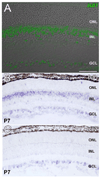
Fig. 1
Fig. 1
The expression of Isl1 in adult mouse retina. Confocal micrograph is overlaid on a phase contrast image of the same retinal section (A). Immunolabeling of Isl1 reveals it is expressed in the INL and GCL but not the ONL. Within the INL, a dense band of immunoreactive cells is present along the outermost INL border, while a more sparse array of immunoreactive cells are found along the innermost INL border, representing putative bipolar and amacrine cell nuclei, respectively. In situ hybridization for Isl1 at P7 demonstrates expression of Isl1 in the INL and GCL (B), while Isl2 expression is restricted to the GCL (C). INL, the inner nuclear layer; GCL, the ganglion cell layer; ONL, the outer nuclear layer. Scale bar = 50 µm.
Fig. 2
Fig. 2
Double labeling of Isl1 and cell-type-specific markers in adult mouse retina. Confocal micrographs of retinal sections co-immunolabeled for Isl1 and various cell-type-specific markers. A,A″: Immunolabeling for Isl1 (A′) and the horizontal cell marker, calbindin (A; arrowheads, asterisks in inset). Isl1 immunoreactivity does not colocalize with calbindin (A″ arrowheads, asterisks in inset). B,B″: Immunolabeling for Isl1 (B′) and the bipolar cell marker, Prox1 (B). Isl1 colocalizes with Prox1 within the outermost portion of the INL (B,B″, larger bracket), whereas along the innermost INL, Isl1 and Prox1 do not colocalize (B,B″, smaller brackets). C,C″: Immunolabeling for Isl1 (C) and the bipolar cell marker, Chx10 (C). Isl1 colocalizes with Chx10 along the outermost INL, whereas Isl1-expressing cells located in the innermost INL and GCL do not express Chx10. D,D″: Immunolabeling for Isl1-LacZ (D′) and the amacrine cell marker, Pax6 (D). Isl1 immunolabeled cells along the innermost INL (arrowheads D,D″) are found within the region of amacrine cells labeled by Pax6 expression. At higher magnification, Pax6 and Isl1 immunoreactivity colocalize (data not shown). The Isl1-lacz reporter gene recapitulates Isl1 immunoreactivity. E,E″: Immunolabeling for Isl1 (E′) and the cholinergic amacrine cell marker, ChAT (E). ChAT-immunoreactive cells are uniformly colocalized with Isl1 (E″). F,F″: Immunolabeling for Isl1 (F′) and the Müller cell marker Kip1 (F) demonstrates that there is no colocalization of Isl1 and Kip1. G,G″: Immunolabeling for Isl1 (G′) and the RGC marker Brn3a (G) demonstrates that a proportion of retinal ganglion cells are immunoreactive for Isl1. Scale bar = 50 µm.
Fig. 2
Fig. 2
Double labeling of Isl1 and cell-type-specific markers in adult mouse retina. Confocal micrographs of retinal sections co-immunolabeled for Isl1 and various cell-type-specific markers. A,A″: Immunolabeling for Isl1 (A′) and the horizontal cell marker, calbindin (A; arrowheads, asterisks in inset). Isl1 immunoreactivity does not colocalize with calbindin (A″ arrowheads, asterisks in inset). B,B″: Immunolabeling for Isl1 (B′) and the bipolar cell marker, Prox1 (B). Isl1 colocalizes with Prox1 within the outermost portion of the INL (B,B″, larger bracket), whereas along the innermost INL, Isl1 and Prox1 do not colocalize (B,B″, smaller brackets). C,C″: Immunolabeling for Isl1 (C) and the bipolar cell marker, Chx10 (C). Isl1 colocalizes with Chx10 along the outermost INL, whereas Isl1-expressing cells located in the innermost INL and GCL do not express Chx10. D,D″: Immunolabeling for Isl1-LacZ (D′) and the amacrine cell marker, Pax6 (D). Isl1 immunolabeled cells along the innermost INL (arrowheads D,D″) are found within the region of amacrine cells labeled by Pax6 expression. At higher magnification, Pax6 and Isl1 immunoreactivity colocalize (data not shown). The Isl1-lacz reporter gene recapitulates Isl1 immunoreactivity. E,E″: Immunolabeling for Isl1 (E′) and the cholinergic amacrine cell marker, ChAT (E). ChAT-immunoreactive cells are uniformly colocalized with Isl1 (E″). F,F″: Immunolabeling for Isl1 (F′) and the Müller cell marker Kip1 (F) demonstrates that there is no colocalization of Isl1 and Kip1. G,G″: Immunolabeling for Isl1 (G′) and the RGC marker Brn3a (G) demonstrates that a proportion of retinal ganglion cells are immunoreactive for Isl1. Scale bar = 50 µm.
Fig. 3
Fig. 3
Double immunolabeling of Isl1 and markers of developing RGCs. Confocal micrographs of horizontal sections of developing mouse retina immunolabeled for Isl1 (A,D) and the developing RGC marker Brn3b (B,E). A–C: Co-immunolabeling for Isl1 and Brn3b at E15.5. Most cells are doubly immunoreactive for Isl1 and Brn3b (brackets in inset), although several cells are singly immunoreactive towards Isl1 (arrowheads in inset). D–F: Co-immunolabeling for Isl1 and Brn3b at E18.5. The distinct array of Isl1 above the band of cells coexpressing both Brn3b and Isl1 represent amacrine cells in the nascent ACL. More cells immunoreactive for only Isl1 are detectable at this stage of development (arrowheads in bracket). Scale bar = 50 µm.
Fig. 4
Fig. 4
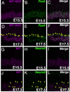
The expression of Isl1 in developing amacrine cells. A–C: Confocal micrographs of a retinal section at E15.5 co-immunolabeled with Isl1-lacZ and Pax6. Cells double-immunopositive for Pax6 and Isl1 are within the ganglion cell layer. D–F: Confocal micrographs of mouse retina at E17.5 co-immunolabeled with Isl1-lacZ and Pax6. The nascent amacrine cell layer is visualized as a distinct lamina of Pax6-expressing cells slightly above the GCL. A proportion of these cells also express Isl1 (arrowheads). G–I: Confocal micrographs of a mouse retinal section at E15.5 co-immunolabeled with Isl1 and NeuroD. Isl1-immunoreactive cells are not colocalized with NeuroD. J–L: Confocal micrographs of a mouse retinal section at E17.5 co-immunolabeled with Isl1 and NeuroD. Several cells immunoreactive for NeuroD in the ganglion and amacrine cell layers (K, arrowheads), also express Isl1 (J,L arrowheads). Scale bar = 50 µm.
Fig. 5
Fig. 5
Isl1 expression in mature bipolar cells. A–C: Confocal micrographs of sections of adult mouse retina co-immunolabeled with Isl1 (A, arrowheads in inset) and the pan-bipolar marker Chx10 (B). Cells immunoreactive for Isl1 are colocalized with Chx10 (C, arrowheads in inset), and are skewed to the upper portion of Chx10-expressing bipolar cells. D–F: Confocal micrographs of sections of adult mouse retina co-immunolabeled with Isl1 and the rod bipolar cell marker PKCα. Cells expressing PKCα (E, arrowheads in inset) also express Isl1 (F, arrowheads in inset). There also exists cells that are PKCα-nonimmunoreactive (E, asterisks in inset) but are Isl1-immunoreactive (F, asterisks in inset). G–I: Confocal micrographs of sections of adult mouse retina co-immunolabeled with Isl1 and the pan-ON-bipolar cell marker Goα. Cells expressing Goα (H, asterisks in inset) also express Isl1 of varying intensities (G,I, asterisks in inset). No cell in this portion of the inner nuclear layer are found to express only Isl1 and not Goα. Scale bar = 50 µm.
Fig. 6
Fig. 6
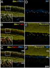
Subtype-specific expression of Isl1 in ON-bipolar cells of mature retina. A–C: Confocal micrographs of sections of adult mouse retina co-immunolabeled for Goα (A–C, asterisks and arrowheads in insets), PKCα (A,C, arrowheads in insets) and Isl1 (B,C, asterisks and arrowheads in insets). Isl1 expression is present in both rod bipolar cells expressing PKCα and Goα (A,C, arrowheads in insets) as well as ON-cone bipolar cells expressing Goα alone (C, asterisks in insets). D–F: Confocal micrographs of sections of adult mouse retina co-immunolabeled for an OFF-cone bipolar marker, recoverin (E,F) and Isl1 (D,F). There is no colocalization between recoverin and Isl1 (F). Scale bar = 50 µm.
Fig. 7
Fig. 7
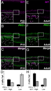
Segregation of Isl1-high expressing bipolar cells with the PKCα-positive rod bipolar subtype. A–C: Confocal micrographs of sections of P10 retina co-immunolabeled for Isl1 and PKCα. Isl1-high expressing cells (A, arrowheads in inset) and Isl1-low expressing cells (A, asterisks in inset) are scored independently and the degree of colocalization of high and low cells with PKCα (B) is scored. PKCα-immunoreactive bipolar cells are more likely to colocalize with a cell expressing relatively higher levels of Isl1 (C, arrowheads vs. asterisks in inset; quantification in D). E–G: Confocal micrographs of sections of adult retina co-immunolabeled for Isl1 and PKCα. Isl1-high expressing cells (E, arrowheads in inset) and Isl1-low expressing cells (E, asterisks in inset) are scored independently, and the degree of colocalization of high and low cells with PKCα (F) is scored. PKCα-immunoreactive cells are more likely to colocalize with a cell expressing relatively higher levels of Isl1 (G, arrowheads vs. asterisks in inset; quantification in H). Scale bar = 50 µm.
Fig. 8
Fig. 8
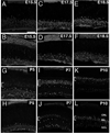
Confocal micrographs of developing mouse retina immunolabeled for Isl1. A,B: Sections of E15.5 retina immunolabeled for Isl1 in central (A) and peripheral (B) retina. Expression of Isl1 is predominant in the developing GCL. C–F: Sections of E17.5 (C,D) and E18.5 (E,F) retina immunolabeled for Isl1 in central (C,E) and peripheral retina (D,F). Expression of Isl1 is maintained in the GCL, but expands into the nascent ACL. This layer corresponds to the distinct lamina of Isl1-expressing cells shown most clearly in central retina (C,E). G,H: Sections of P5 retina immunolabeled for Isl1 in central (G) and peripheral (H) retina. Expression of Isl1 in the presumptive bipolar region of the INL commences in central retina at this stage (G, bracket), while peripheral retina is devoid of Isl1 immunolabeling in this region. I–L: Sections of P7 (I–L) and P10 (K,L) retinas immunolabeled for Isl1 in central (I,K) and peripheral retina (J,L). The expression of Isl1 in the presumptive bipolar cell layer (I–L, brackets) continues to expand into peripheral retina (J,L, brackets). Note the elongated nuclei in recently migrated bipolar cells in central retina at P7 (I, brackets) and peripheral retina at P7 and P10 (J,L brackets; respectively). Scale bar = 50 µm.
Fig. 9
Fig. 9
Coexpression of Isl1 and Chx10 in developing bipolar cells. A–C: Confocal micrographs of sections of P5 central retina co-immunolabeled for Isl1 (A) and Chx10 (B). The nascent Isl1 expression in presumptive bipolar cells (A, bracket) spatially overlaps with the domain of Chx10 expression (C). D–F: Confocal micrographs of sections of P7 central retina co-immunolabeled for Isl1 (D) and Chx10 (E). Colocalization of Isl1 and Chx10 at this stage shows that double immunopositive cells (F, white) are randomly interspersed throughout the pool of developing bipolar cells. G–I: Confocal micrographs of sections of P10 central retina co-immunolabeled for Isl1 (G) and Chx10 (H). Colocalization of Isl1 and Chx10 is most notable at the upper boundary of Chx10 expression domain. Note the transition of coexpressing cells from a random assortment in P7 (F) to a lamina (I) of presumptive ON-bipolar cells. Scale bar = 50 µm.

Similar articles

Cited by

References

    1. Beleky-Adams T, Tomarev S, Li HS, Ploder L, McInnes RR, Sundin O, Adler R. Pax-6, Prox 1, and Chx10 homeobox gene expression correlates with phenotypic fate of retinal precursor cells. Invest Ophthalmol Vis Sci. 1997;38:1293–1303. - PubMed
    1. Bramblett DE, Pennesi ME, Wu SM, Tsai MJ. The transcription factor Bhlhb4 is required for rod bipolar cell maturation. Neuron. 2004;43:779–793. - PubMed
    1. Brown SP, Masland RH. Costratification of a population of bipolar cells with the direction-selective circuitry of the rabbit retina. J Comp Neurol. 1999;408:97–106. - PubMed
    1. Brunelli G, Spano P, Barlati S, Guarneri B, Barbon A, Bresciani R, Pizzi M. Glutamatergic reinnervation through peripheral nerve graft dictates assembly of glutamatergic synapses at rat skeletal muscle. Proc Natl Acad Sci USA. 2005;102:8752–8757. - PMC - PubMed
    1. Cepko CL, Austin CP, Yang X, Alexiades M, Ezzeddine D. Cell fate determination in the vertebrate retina. Proc Natl Acad Sci USA. 1996;93:589–595. - PMC - PubMed

Publication types

MeSH terms

Substances